. 2007 Mar 28;27(13):3416-28.
doi: 10.1523/JNEUROSCI.0273-07.2007.

Myelination of congenitally dysmyelinated spinal cord axons by adult neural precursor cells results in formation of nodes of Ranvier and improved axonal conduction

Affiliations

Myelination of congenitally dysmyelinated spinal cord axons by adult neural precursor cells results in formation of nodes of Ranvier and improved axonal conduction

Eftekhar Eftekharpour et al. J Neurosci. .

Abstract

Emerging evidence suggests that cell-based remyelination strategies may be a feasible therapeutic approach for CNS diseases characterized by myelin deficiency as a result of trauma, congenital anomalies, or diseases. Although experimental demyelination models targeted at the transient elimination of oligodendrocytes have suggested that transplantation-based remyelination can partially restore axonal molecular structure and function, it is not clear whether such therapeutic approaches can be used to achieve functional remyelination in models associated with long-term, irreversible myelin deficiency. In this study, we transplanted adult neural precursor cells (aNPCs) from the brain of adult transgenic mice into the spinal cords of adult Shiverer (shi/shi) mice, which lack compact CNS myelin. Six weeks after transplantation, the transplanted aNPCs expressed oligodendrocyte markers, including MBP, migrated extensively along the white matter tracts of the spinal cord, and formed compact myelin. Conventional and three-dimensional confocal and electron microscopy revealed axonal ensheathment, establishment of paranodal junctional complexes leading to de novo formation of nodes of Ranvier, and partial reconstruction of the juxtaparanodal and paranodal molecular regions of axons based on Kv1.2 and Caspr (contactin-associated protein) expression by the transplanted aNPCs. Electrophysiological recordings revealed improved axonal conduction along the transplanted segments of spinal cords. We conclude that myelination of congenitally dysmyelinated adult CNS axons by grafted aNPCs results in the formation of compact myelin, reconstruction of nodes of Ranvier, and enhanced axonal conduction. These data suggest the therapeutic potential of aNPCs to promote functionally significant myelination in CNS disorders characterized by longstanding myelin deficiency.


Figures

Figure 1.
Figure 1.
Multipotential ability of aNPCs to generate neural cells in vitro. YFP-derived neurospheres (A–C) from the subventricular zone of adult transgenic mice expressed nestin. After 2–3 passages, the cells were dissociated and then cultured as a monolayer on matrigel-coated multichamber glass slides. For the first 2 d, they were maintained in serum-free growth medium containing EGF and bFGF. Immunocytochemistry on these cultures, 48 h after plating, showed uniform bipolar cells that were positive for nestin (D–F). The nestin-positive stem/progenitor cells were switched to a serum-containing medium. Five days later, immunostaining for neuronal and glial markers showed differentiation toward astrocytes (G–I), oligodendroglia (J–O), and neurons (P–R). Although the majority of YFP-derived aNPCs differentiated into GFAP-positive astrocytes in vitro (G–I), we also observed CNPase-positive mature oligodendrocytes (J–L), PDGF-αR-positive immature oligodendroglia (M–O), and MAP-2-positive mature neurons (P–R).
Figure 2.
Figure 2.
YFP-labeled aNPCs survive, integrate, and migrate along white matter tracts in shi/shi spinal cord. A, Confocal image of a longitudinal section of transplanted shi/shi spinal cord. Six weeks after transplantation, YFP-labeled aNPCs migrated up to 2–3 mm away from the injection sites (*) along the rostrocaudal axis of the spinal cord. B, Immunostaining for the neuronal marker βIII-tubulin (Tuj1) showed the tendency of YFP-derived cells to reside predominantly in the white matter. C, Higher magnification of YFP-derived cells with extended processes along the white matter tracts.
Figure 3.
Figure 3.
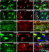
YFP-positive aNPCs differentiate principally into glial lineage in vivo. Confocal immunohistochemistry of spinal cord sections from transplanted shi/shi mice showed that the majority of YFP-labeled aNPCs expressed olig2, an oligodendroglial transcription factor, 6 weeks after transplantation (A–C). A significant proportion of YFP-derived cells showed colocalization of YFP-positive cells with PDGF-αR, a marker for immature oligodendrocytes (D–F). We also observed immunoreactivity for mature oligodendrocytes as shown by APC labeling (G–L). Our quantitative analysis on three transplanted shi/shi mice at 6 weeks after transplantation showed that ∼78 ± 9% of YFP-positive cells expressed olig2, which is expressed in oligodendrocytes at all developmental stages. To further characterize the extent of differentiation of the aNPCs into oligodendroglial cells, we found that 43 ± 7% expressed APC and 15 ± 5% expressed PDGF-αR. A small proportion of YFP cells (9% ± 4) also showed GFAP immunoreactivity in their cell bodies and were thus considered as astrocytes (M–O).
Figure 4.
Figure 4.
YFP-positive aNPCs do not express nestin or markers of neurons or Schwann cells. Confocal immunohistochemistry of transplanted shi/shi mice 6 weeks after transplantation showed no nestin expression in aNPCs (A–C). Although some of the YFP-labeled aNPCs were closely associated with Tuj1-positive neuronal processes (D–E), no YFP-positive neuronal somata were observed. We also did not detect any p75 (G–I) or myelinating P0-positive (J–L) Schwann cells among YFP-positive aNPCs. Insets in I and L show control positive staining for p75 and P0 in the peripheral roots of the shi/shi spinal cord.
Figure 5.
Figure 5.
YFP-positive oligodendrocytes express MBP. Confocal immunohistochemistry on transplanted shi/shi mice spinal cord sections revealed MBP expression by transplanted aNPCs. Although no MBP was detectable in the naive shi/shi spinal cords, transplanted spinal cord segments of shi/shi mice showed patches of MBP as early as 1 week after transplantation (A–C). After 6 weeks, NPC-transplanted segments displayed extensive MBP expression (D–F). After NPC transplantation, one YFP-positive oligodendrocyte ensheathed several surrounding axons (labeled with NF200) and produced MBP (G–N). Three-dimensional reconstruction of YFP-positive transplanted cells in close association with the host spinal cord axons is shown (O).
Figure 6.
Figure 6.
NPC transplants induced formation of compact myelin, appearance of nodes of Ranvier, and paranodal junctional complex. Electron micrographs from a 14-week-old naive shi/shi spinal cord (A) showed a lack of compact myelin. The myelin sheaths in shi/shi mice were characterized by 2–3 layers of noncompacted membrane (B). Six weeks after transplantation, many myelinated axons were observed in transplanted segments of spinal cord axons (C, D) containing multilayered compact myelin (E, F). Electron microscopy on longitudinal sections of spinal cord in wild type (G), naive shi/shi (H), and transplanted (I) to show the paranodal junctional complex. Paranodal junctions (arrows) were detectable on either side of the nodes of Ranvier in wild-type animals (G) marking the paranodal zone. We did not recognize the paranodal zone in control animals, and only sporadic and irregular contacts between the glial cells and axonal membrane were detected (H, arrows). Occasionally, node-like structures were observed in transplanted animals (I). Higher-magnification micrographs confirmed the presence of paranodal zones in which the processes of transplanted aNPC-derived myelinating oligodendrocytes formed paranodal junctional complex with the axonal membrane (J, K, arrows).
Figure 7.
Figure 7.
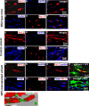
Transplanted aNPCs promote the aggregation of K+ channels and the formation of nodes of Ranvier in the spinal cord axons of shi/shi mice. Confocal immunostaining of Kv1.2 subunits (red) and pan-Na+ channels (blue) in the spinal cord of wild-type mice (A–C), control shi/shi mice (G–I), and transplanted shi/shi mice (M–P) is depicted. Kv1.2 subunits were clearly localized to the juxtaparanodal regions of wild-type spinal cord axons (A–C), confirmed with nodal pan-Na+ immunostaining. In shi/shi mice, Kv1.2 immunostaining was abnormally distributed along the axonal internodes (G–I), but Na+ clusters were observed as aberrant nodal aggregates. Six weeks after aNPCs transplantation, spinal cord segments of shi/shi mice showed restoration of Kv1.2 subunit clusters (M–P). YFP-positive processes of transplanted aNPCs were observed in close association with axons containing restored K+ channels aggregates (P). Nodal localization of Na+ channels was further confirmed using Nav1.6 (red) immunostaining in wild-type (D–F), control shi/shi (J–L), and transplanted shi/shi (Q–T). Caspr (blue) immunostaining was used to identify the paranodal area. A 3D reconstruction clearly shows a node of Ranvier that is bordered by an MBP-expressing NPC derived oligodendrocyte. Note that the processes of YFP-labeled oligodendrocytes avoid the nodal region (U).
Figure 8.
Figure 8.
Transplantation of aNPCs leads to shortened distribution of K+ channel and Caspr and reconstruction of paranodal and juxtaparanodal regions in the spinal cord axons of shi/shi mice. The normal juxtaparanodal localization of Kv1.2 channel subunits and the paranodal localization of Caspr (A–F) in wild-type mice were completely disrupted in the spinal cord axons of control shi/shi mice (G–L). In contrast, the transplanted segments of shi/shi mice spinal cords showed evidence of a more normal distribution of Kv1.2 channel subunit and Caspr (M–R). Quantification of the confocal immunostaining data revealed that the distribution of the Kv1.2 K+ channel subunits in the transplanted area was significantly more compact (70%) than in the segments of the spinal cord of the same mice, which was remote to the site of transplantation. Of note, Caspr immunohistochemistry also showed a significant (27%) decrease in the longitudinal distribution along the axons. Box plots S and T show the distribution of total length of Kv1.2 and Caspr in all three groups (Kruskal–Wallis one-way ANOVA on ranks, *p < 0.001; n = 4 per group).
Figure 9.
Figure 9.
Transplantation of aNPCs improves axonal conduction in shi/shi mouse as shown by increased amplitude and decreased latency. A, Electrodes were positioned 5 mm apart on the dorsal surface of the cord at the transplanted area. Control recordings were done from either nontransplanted segments (T10–L1) of the transplanted animal or from thoracic segments of control (nontransplanted) shi/shi mice: B, wild-type mouse; C, control shi/shi spinal cord; D, nontransplanted segments of cord; and E, transplanted shi/shi mouse cord. F, At the end of the experiment, the cord was cut to confirm the specificity of the CAP. Electrophysiological measurements showed reduced latency, increased amplitude, and enhanced estimated conduction velocity of transplanted spinal cord segments when compared with nontransplanted cord segments (paired t test, *p = 0.04; n = 5) in transplanted spinal cords.

Comment in

References

    1. Arroyo EJ, Xu T, Grinspan J, Lambert S, Levinson SR, Brophy PJ, Peles E, Scherer SS. Genetic dysmyelination alters the molecular architecture of the nodal region. J Neurosci. 2002;22:1726–1737. - PMC - PubMed
    1. Baba H, Akita H, Ishibashi T, Inoue Y, Nakahira K, Ikenaka K. Completion of myelin compaction, but not the attachment of oligodendroglial processes triggers K+-channel clustering. J Neurosci Res. 1999;58:752–764. - PubMed
    1. Baron-Van Evercooren A, Clerin-Duhamel E, Lapie P, Gansmuller A, Lachapelle F, Gumpel M. The fate of Schwann cells transplanted in the brain during development. Dev Neurosci. 1992;14:73–84. - PubMed
    1. Bird TD, Farrell DF, Sumi SM. Brain lipid composition of the shiverer mouse (genetic defect in myelin development) J Neurochem. 1978;31:387–391. - PubMed
    1. Black JA, Waxman SG, Smith KJ. Remyelination of dorsal column axons by endogenous Schwann cells restores the normal pattern of Nav1.6 and Kv1.2 at nodes of Ranvier. Brain. 2006;129:1319–1329. - PubMed

Publication types

MeSH terms

Substances