Clinical Trial
. 2007 Mar 28;27(13):3429-38.
doi: 10.1523/JNEUROSCI.4273-06.2007.

Functional coupling of human prefrontal and premotor areas during cognitive manipulation

Affiliations
Clinical Trial

Functional coupling of human prefrontal and premotor areas during cognitive manipulation

Mitsunari Abe et al. J Neurosci. .

Abstract

Evidence indicates the involvement of the rostral part of the dorsal premotor cortex (pre-PMd) in executive processes during working memory tasks. However, it remains unclear what the executive function of pre-PMd is in relation to that of the dorsolateral prefrontal cortex (DLPFC) and how these two areas interact. Using functional magnetic resonance imaging (fMRI), brain activity was examined during a delayed-encoding recognition task. Fifteen subjects had prelearned several four-code standard sequences and super sequences (SUPs) consisting of a train of two standard sequences to form "chunks" in long-term memory. During fMRI, subjects remembered eight-code encoding stimuli presented as an SUP or two unlinked standard sequences (2STs). A memory probe prompted the subjects to recognize codes across two chunks (ACROSS) or within a single chunk. A 2 x 2 factorial design was used to test two types of working memory manipulation: (1) a reductive operation selecting codes from chunks ("segmenting") and (2) a synthetic operation converting unlinked codes into a sequence ("binding"). Response time data supported the behavioral effects of each operation. Event-related fMRI showed that the "segmenting operation" activated the DLPFC bilaterally, whereas the "binding operation" enhanced the left pre-PMd activity. Activity in the ventrolateral prefrontal cortex suggested its involvement in the retrieval of task-relevant information from long-term memory. Furthermore, effective connectivity analysis indicated that the left pre-PMd and ipsilateral DLPFC interacted specifically during the ACROSS recognition of 2STs, the condition that involved both operations. We propose specific neural substrates for working memory manipulation: the DLPFC for segmenting/attentional selection and the pre-PMd for binding/sequencing. The functional coupling between the DLPFC and pre-PMd appears to play a role in combining these distinct operations.


Figures
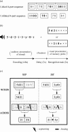
Figure 1.
Figure 1.
Experimental procedures. a, Background of the chunk-constrained manipulation of memory. Imagine registering a familiar telephone number represented in WM. This 11-bit example sequence is typically recoded into a single four-part sequence (top, linked 4-part sequence). The phone number sequence can be regarded as a multilevel memory chunk in LTM. However, if this sequence was rearranged in the reversed order, then the linkage between the elements would be broken between the four chunks, whereas it would be preserved within each chunk (bottom, unlinked 4-part sequence). In both sequences, the retrieval of codes extending over two chunks (e.g., 5136 or 0375) requires more demanding behavioral cost than that of the same number of codes already forming a chunk (e.g., 3603). This process requires one to select parts of the chunks (segmenting). Moreover, in the reversed sequence, these segmented parts should be forming a new sequence (binding), because the linkages between the adjacent parts had been broken through the rearrangement. These processes, segmenting and binding, conceivably occur within WM, and the segmenting requires the central executive to not only operate on WM contents but also to access to the previous knowledge of chunk structures in LTM. b, In the fMRI experiment, a trial started with an auditory presentation of eight-code encoding stimuli as an SUP or a train of 2ST for 4.8 s, followed by a maintenance period during which a fixation cross was presented for 3 s. A memory probe either for WITHIN recognition or ACROSS recognition was visually presented for 2 s. In the example, the memory probe for the WITHIN recognition (the top probe) matched with the corresponding part of the encoding sequence (match), whereas the probe for the ACROSS recognition (the bottom probe) did not (mismatch). The intertrial interval was 15 s. c, Schema representing possible cognitive processes in the four recognition conditions in a 2 × 2 factorial design. It illustrates presumable recoding–recognition patterns of an SUP stimulus (5d2i2h5f, left panels in the 2 × 2 grids) and of a 2ST stimulus (5d2i3y8r, right panels). Compared with the WITHIN recognition, ACROSS recognition should require greater attentional selection or more demanding segmenting operations (split-gap). The ACROSS recognition of 2ST stimuli should more rigorously demand the binding operation than the other recognition conditions (double line).
Figure 2.
Figure 2.
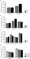
Response time analysis. a, Mean response times in the four recognition conditions. b, Reanalysis of the response time data, taking the three positions of the memory probe into account. c, Reanalysis of the response times in the four recognition conditions, taking the case effect (match or mismatch) into account. AC, ACROSS recognition; WI, WITHIN recognition. d, Time-dependent changes in response times during the fMRI experiment (STAGE effect). An fMRI session including 72 trials was split into three stages. Error bars indicate SEM.
Figure 3.
Figure 3.
a, Recoding-related activity superimposed onto a surface-rendered standard brain. White circle, Left VLPFC; yellow circle, left frontal operculum; orange circle, pre-SMA; green circle, left IPL. b, The recoding regions are shown on three slices of the mean anatomical MRI from 15 subjects. An axial view (z = 38 mm) showing activities in the left frontal operculum (yellow circle) and the left IPL (green circle) network. An axial view (z = 59 mm) shows pre-SMA activity (orange circle). A coronal view (y = 32 mm) shows activity in the left VLPFC (white circle). Axial and sagittal views (inset, gray dashed line box) show that the recoding-related VLPFC activity (blue) was adjacent to the segmenting-related VLPFC activity (red). Lt, Left; Rt, right.
Figure 4.
Figure 4.
a, Segmenting-related activity superimposed onto a surface-rendered standard brain. b, Activity was overlaid onto slices of the averaged anatomical MRI from 15 subjects. An axial view (z = 25 mm) shows activity in the left DLPFC in a green circle. A coronal view (y = 25 mm) shows activity in the right DLPFC (green circle) and bilateral VLPFC (white circles). An axial view (z = 45 mm) shows activity in the bilateral superior parietal lobules (blue circles). Lt, Left; Rt, right. c, Left DLPFC activity (percentage of signal changes) is plotted for the four conditions. Error bars indicate SEM.
Figure 5.
Figure 5.
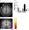
a, Binding-related activity overlaid onto slices (top, z = 53 mm; bottom, y = 3 mm) of the averaged anatomical MRI. Activation is located in the depth of the SFS (white dashed arrow) and anterior to the superior precentral sulcus (white arrow). b, Left pre-PMd activity (percentage of signal changes) is plotted for the four conditions. The pre-PMd activity was significantly greater than the baseline only during the 2ST-ACROSS condition in the whole-brain corrected analysis (FDR threshold of p = 0.05). Error bars indicate SEM. Lt, Left; Rt, right.
Figure 6.
Figure 6.
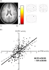
a, Significant activity obtained from PPI analysis is superimposed onto an axial slice (z = 23 mm) of the averaged anatomical MRI (left). Only the left DLPFC (x = −40; y = 24; z = 23) exhibits a significant PPI effect (z value = 3.47) at a liberal threshold of uncorrected p < 0.01 (right). Lt, Left; Rt, right. b, Data from a representative subject showing the relationship between left pre-PMd activity (x-axis) and left DLPFC activity (y-axis) during SUP-ACROSS and 2ST-ACROSS. Regression lines are shown as a gray dashed line for the SUP-ACROSS condition and a black line for the 2ST-ACROSS condition. The crosses (SUP) and squares (SUB) represent adjusted signals extracted from all observed scans during the two ACROSS conditions (156 data points each).

References

    1. Baddeley A. The episodic buffer: a new component of working memory? Trends Cogn Sci. 2000;4:417–423. - PubMed
    1. Barbas H, Pandya DN. Architecture and frontal cortical connections of the premotor cortex (area 6) in the rhesus monkey. J Comp Neurol. 1987;256:211–228. - PubMed
    1. Bor D, Cumming N, Scott CE, Owen AM. Prefrontal cortical involvement in verbal encoding strategies. Eur J Neurosci. 2004;19:3365–3370. - PubMed
    1. Braver TS, Cohen JD, Nystrom LE, Jonides J, Smith EE, Noll DC. A parametric study of prefrontal cortex involvement in human working memory. NeuroImage. 1997;5:49–62. - PubMed
    1. Callicott JH, Mattay VS, Bertolino A, Finn K, Coppola R, Frank JA, Goldberg TE, Weinberger DR. Physiological characteristics of capacity constraints in working memory as revealed by functional MRI. Cereb Cortex. 1999;9:20–26. - PubMed

Publication types