. 2007 Jun;18(6):2102-11.
doi: 10.1091/mbc.e06-12-1075. Epub 2007 Mar 28.

TRIM5 alpha cytoplasmic bodies are highly dynamic structures

Affiliations

TRIM5 alpha cytoplasmic bodies are highly dynamic structures

Edward M Campbell et al. Mol Biol Cell. 2007 Jun.

Abstract

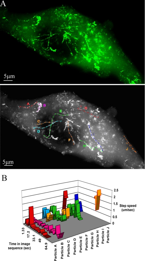
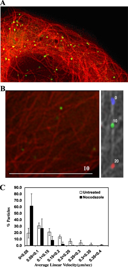
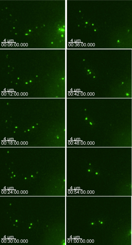
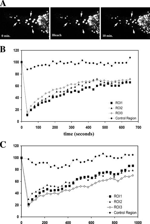
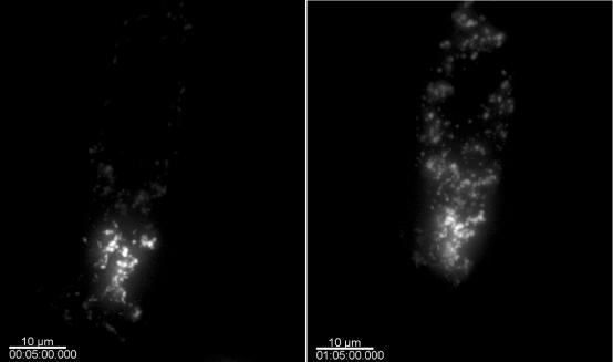
Tripartite motif (TRIM)5 alpha has recently been identified as a host restriction factor that has the ability to block infection by certain retroviruses in a species-dependent manner. One interesting feature of this protein is that it is localized in distinct cytoplasmic clusters designated as cytoplasmic bodies. The potential role of these cytoplasmic bodies in TRIM5 alpha function remains to be defined. By using fluorescent fusion proteins and live cell microscopy, we studied the localization and dynamics of TRIM5 alpha cytoplasmic bodies. This analysis reveals that cytoplasmic bodies are highly mobile, exhibiting both short saltatory movements and unidirectional long-distance movements along the microtubule network. The morphology of the cytoplasmic bodies is also dynamic. Finally, photobleaching and photoactivation analysis reveals that the TRIM5 alpha protein present in the cytoplasmic bodies is very dynamic, rapidly exchanging between cytoplasmic bodies and a more diffuse cytoplasmic population. Therefore, TRIM5 alpha cytoplasmic bodies are dynamic structures more consistent with a role in function or regulation rather than protein aggregates or inclusion bodies that represent dead-end static structures.


Figures

Figure 1.
Figure 1.
Functional validation of YFP-TRIM5α fusion proteins and the cellular localization of TRIM5α fusion proteins. Stable cell lines expressing YFP-TRIM5α fusion proteins were tested for their ability to restrict HIV-1 (A) or SIV (B) reporter viruses. YFP-rhTRIM5α (squares) effectively inhibited HIV-1 infection relative to HeLa control (circles), but it less effectively inhibited SIV infection. YFP-huTRIM5α (triangles) expression only minimally inhibited HIV-1 infection. (C) 293T cells were transfected with YFP-TRIM5α fusion proteins. YFP-huTRIM5α and YFP-rhTRIM5α fusion proteins were expressed as intact fusions when compared with unfused YFP.
Figure 2.
Figure 2.
(A) HeLa cells were transiently transfected with YFP-huTRIM5α, and the cells then fixed, stained for cellular DNA (blue), and TRIM5α localization (green) was examined. An enlargement highlighting the hollow body structures formed by YFP-hu-TRIM5α is shown in the top inset. (B) Electron microscopy confirms that YFP-huTRIM5α forms hollow structures, with a magnification of the structures observed shown in the bottom inset. The P-body markers mRFP-Dcp1a (C) and RFP-rck/p54 were expressed in cells stably expressing YFP-rhTRIM5α. Cells were fixed and analyzed by confocal microscopy. The merged images demonstrates the lack of TRIM5α (green) and Dcp1a (red) (C) or RFP-rck/p54 (red) (D) colocalization.
Figure 3.
Figure 3.
TRIM5α particle movement and determination of the movement rates. (A) A live TE671 cell stably expressing GFP-huTRIM5α was imaged over 60 frames at 1.325-s intervals (0.75 frames/s). The 60 individual frames were summed using ImageJ on the basis that in the composite image, a given pixel is assigned the maximum gray scale value (0-256) observed in any of the 60 frames throughout the series. The tracks of a number of cytoplasmic bodies are shown in the top panel. The directionality and authenticity of these tracks were confirmed by comparison with Supplemental Movie 1. Several prominent tracks were identified for further analysis. These tracks are labeled A–J in bottom panel. (B) Velocities of particles A–J in A were measured throughout the time that they were observed during the 60-frame (80-s) sequence by using the manual tracking function of the particle tracking software GMimPro. The vertical y-axis shows the average speed of the particle between each consecutive frame (step speed; micrometers per second); the horizontal-z-axis shows the relative time during the image sequence in which the particular particle was observed.
Figure 4.
Figure 4.
Movement of individual cytoplasmic bodies upon microtubules and reduction in cytoplasmic body motility upon treatment with nocodazole. (A) A static image demonstrating the association of GFP-huTRIM5α (green) and CFP-labeled tubulin (red). (B) A live TE671 cell stably expressing CFP-labeled tubulin and GFP-huTRIM5α was imaged using fluorescence microscopy. Left, starting positions for a cytoplasmic body (green) and the microtubule network (red). Right, microtubule network in gray with the relative positions of the cytoplasmic body at 10-s intervals colored blue, green, and red. The relative time (seconds) is noted by the body. (C) The movement of GFP-TRIM5α stably expressed in TE671 cells was monitored by live cell microscopy. Cells were either left untreated or treated with 66 μM nocodazole for 2 h before imaging. Images were captured at a rate of 0.75 frames per second for a total of 60 frames. Particle tracks were analyzed using GMview, and they were verified manually to confirm authenticity. Five randomly chosen cells were analyzed in each case, resulting in 258 cytoplasmic body tracks for untreated cells and 306 tracks for nocodazole-treated cells. Velocities were grouped into 0.05 μm/s bins for each cell analyzed, and the percentage of particles in each bin was determined. The mean and SD values in each bin over five cells were then calculated.
Figure 5.
Figure 5.
Smaller YFP TRIM5α cytoplasmic bodies combine to form larger bodies. HOS cells were microinjected with GFP-rhTRIM5α, and cells were imaged at 2-min intervals. At the onset of expression, small cytoplasmic bodies quickly became apparent, and these smaller bodies could combine to form larger bodies.
Figure 6.
Figure 6.
FRAP analysis of TRIM5α cytoplasmic bodies. (A) Individual YFP-TRIM5α bodies were photobleached using a confocal laser, and fluorescence of the bleached body was monitored over time. Recovery of YFP-huTRIM5α (B) was similar to recovery observed in rhYFP-TRIM5α bodies (C).
Figure 7.
Figure 7.
Photoactivation analysis of TRIM5α cytoplasmic bodies. A HeLa cell transiently transfected with PAGFP-TRIM5α is activated in a discreet region, whereas the rest of the PAGFP-TRIM5α remains unactivated and therefore not fluorescent. TRIM5α dynamics is examined by monitoring the behavior of this fluorescent subpopulation of PAGFP-TRIM5α. A cell immediately after photoactivation (left) and 60 min later (right) is shown.

References

    1. Anderson P., Kedersha N. RNA granules. J. Cell Biol. 2006;172:803–808. - PMC - PubMed
    1. Best S., Le Tissier P., Towers G., Stoye J. P. Positional cloning of the mouse retrovirus restriction gene Fv1. Nature. 1996;382:826–829. - PubMed
    1. Bieniasz P. D. Intrinsic immunity: a front-line defense against viral attack. Nat. Immunol. 2004;5:1109–1115. - PubMed
    1. Campbell E. M., Nunez R., Hope T. J. Disruption of the actin cytoskeleton can complement the ability of Nef to enhance human immunodeficiency virus type 1 infectivity. J. Virol. 2004;78:5745–5755. - PMC - PubMed
    1. Cohen P. Making a monkey out of HIV. More is becoming clear about a novel host factor that appears central to governing the species-specificity of retroviruses like HIV and could be a future antiviral target. IAVI Rep. 2005;9:9–12. - PubMed

Publication types

MeSH terms

LinkOut - more resources