

Brown-Sequard syndrome after blunt cervical spine trauma: clinical and radiological correlations
- PMID: 17394028
- PMCID: PMC2200771
- DOI: 10.1007/s00586-007-0345-7
Brown-Sequard syndrome after blunt cervical spine trauma: clinical and radiological correlations
Abstract
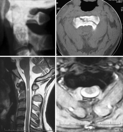
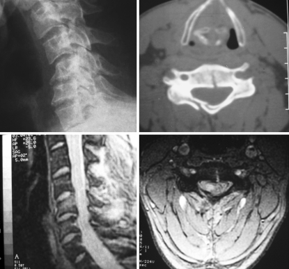
The objective of this study was to describe clinical and radiological features of a series of patients presenting with Brown-Sequard syndrome after blunt spinal trauma and to determine whether a correlation exists between cervical plain films, CT, MRI and the clinical presentation and neurological outcome. A retrospective review was done of the medical records and analysis of clinical and radiological features of patients diagnosed of BSS after blunt cervical spine trauma and admitted to our hospital between 1995 and 2005. Ten patients were collected for study, three with upper- and seven with lower-cervical spine fracture. ASIA impairment scale and motor score were determined on admission and at last follow-up (6 months-9 years, mean 30 months). Patients with lower cervical spine fracture presented with laminar fracture ipsilateral to the side of cord injury in five out of six cases. T2-weighted hyperintensity was present in seven patients showing a close correlation with neurological deficit in terms of side and level but not with the severity of motor deficit. Patients with Brown-Sequard syndrome secondary to blunt cervical spine injury commonly presented T2-weighted hyperintensity in the clinically affected hemicord. A close correlation was observed between these signal changes in the MR studies and the neurologic level. Effacement of the anterior cervical subarachnoid space was present in all patients, standing as a highly sensitive but very nonspecific finding. In the present study, craniocaudal extent of T2-weighted hyperintensity of the cord failed to demonstrate a positive correlation with neurological impairment.
Figures
References
-
- Berlit P. Rare causes of the Brown-Sequard syndrome. NMR tomographic findings. Dtsch Med Wochenschr. 1988;113:1844–1846. - PubMed
-
- Brown-Sequard CE (1973): De la trasmission des impressions sensitives par la moelle epiniere-CR Soc Biol 1849:1:192–194. In: Wilkins RH, Brody IA, (eds) English translation in neurological classics. Johnson Reprint Corporation, New York, pp 49–50
-
- Collignon F, Martin D, Lenelle J, Stevenaert A. Acute traumatic central cord syndrome: magnetic resonance imaging and clinical observations. J Neurosurg. 2002;96(1 Suppl):29–33. - PubMed
MeSH terms
LinkOut - more resources
Full Text Sources
Medical
