. 2007 Apr 24;23(9):5045-9.
doi: 10.1021/la063765e. Epub 2007 Mar 30.

Multiscale structure of the underwater adhesive of Phragmatopoma californica: a nanostructured latex with a steep microporosity gradient

Affiliations

Multiscale structure of the underwater adhesive of Phragmatopoma californica: a nanostructured latex with a steep microporosity gradient

Mark J Stevens et al. Langmuir. .

Abstract

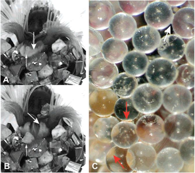
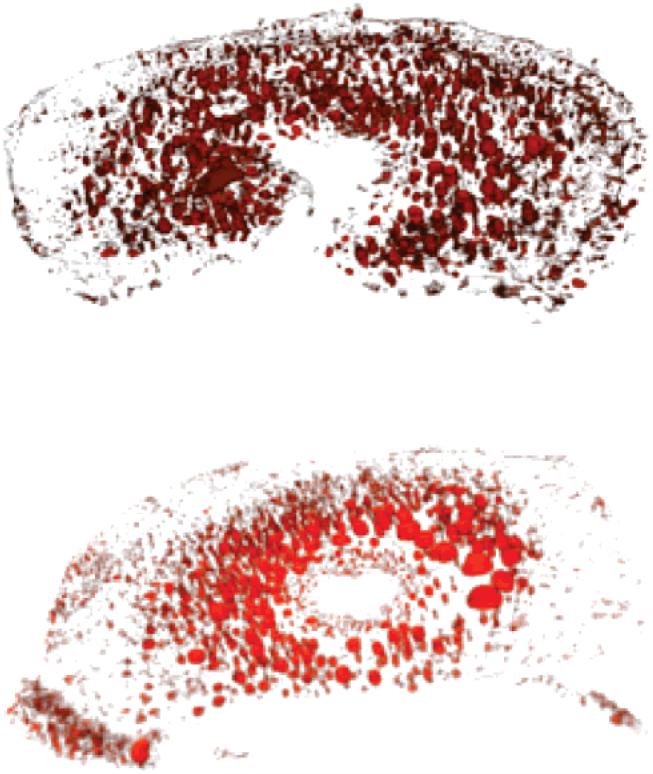
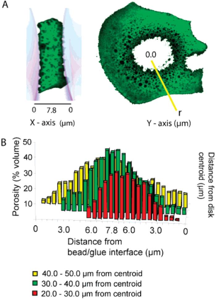
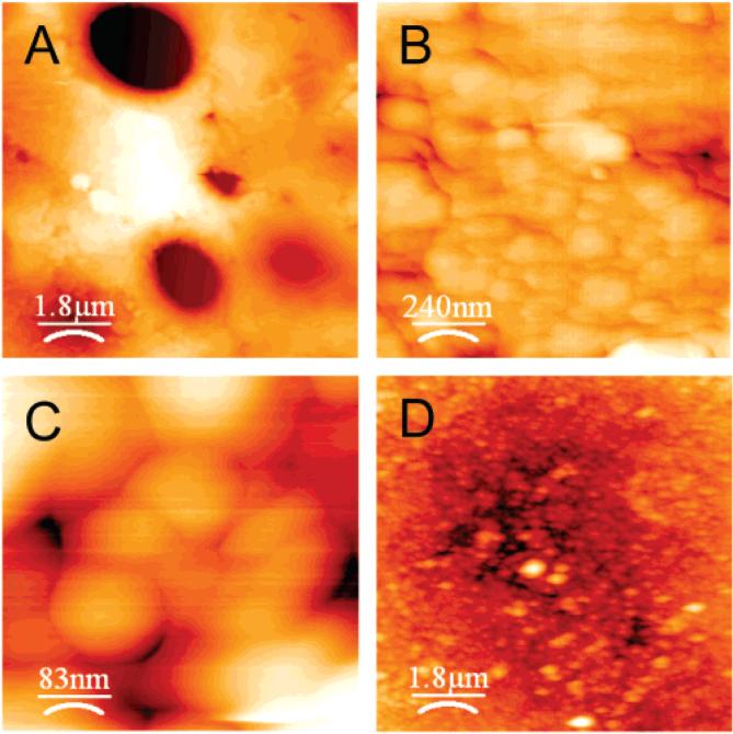
Phragmatopoma Californica builds a tubular dwelling by gluing bits of sand and seashell together underwater with a proteinaceous adhesive. In the lab, the animals will build with 0.5 mm glass beads. Two spots of glue with a consistent volume of about 100 pL each are deposited on the glass beads before placement on the end of the tube. The animals wriggled the particles for 20-30 s before letting go, which suggested that the adhesive was sufficiently set within 30 s to support the glass beads. The structure of the adhesive joints was examined at the micro- and nanoscopic length scales using laser scanning confocal and atomic force microscopies. At the microscale, the adhesive was a cellular solid with cell diameters ranging from 0.5 to 6.0 mum, distributed to create a steep porosity gradient that ranged from near zero at the outside edges to about 50% at the center of the adhesive joint. At the nanoscale, the adhesive appeared to be an accretion of trillions of deformable nanospheres, reminiscent of a high-solids-content latex adhesive. The implications of the structure for the functionality of the adhesive is discussed.


Figures

Figure 1
Figure 1
P. californica adding a sand grain to its tube. (A) After applying dabs of glue, P. californica nestles a sand grain into place and holds it for 20-30 s before letting go. (B) Building organ. Large arrows indicates the building organ after release of a sand grain. Small arrows indicate the freshly adhered sand grain. (C) When freshly deposited, the glue is creamy white (white arrow). After several hours, the glue turns rusty brown in color (red arrows).
Figure 2
Figure 2
Porous volume of two representative glue disks rendered in three dimensions by reconstructing laser scanning confocal microscopy optical sections. The nearly solid edges and higher porous central regions are evident.
Figure 3
Figure 3
Porosity (Z-axis) measured as a function of distance from glue-substrate interface (X-axis) and as a function of distance (r) from the centroid of the glue disk (Y-axis). (A) Representative confocal fluorescent images of glue disks and definition of porosity plot axes. (B) Porosity varies as a function of distance from the glue/bead interface and as a function of radial distance (r) from the centroid. For clarity error bars were not added to the bar graph; errors in all cases were less than 10%.
Figure 4
Figure 4
Representative AFM images of P.californica glue. (A-C) Dried and cohesively fractured glue disks at increasing resolution. (D) Hydrated glue disk.
Figure 5
Figure 5
P. californica adhesive model. Within secretory cells of the cement glands a mixture of the oppositely charged adhesive proteins (Pc1-3) and divalent cations condense into a nanoparticulate fluid phase through complex coacervation. Initial adhesion between the deformable nanoparticles may be mediated by electrostatic bridging of excess negative surface charges by divalent cations and strand exchange between particles. When secreted, the high-solids-content adhesive latex is denser than seawater, has sufficient viscosity and cohesion to prevent rapid dissolution into the seawater, but low interfacial tension to allow spreading and crack filling on the wet mineral substrate. A discontinuous aqueous phase trapped in the setting adhesive forms a microporous foam structure. A two-stage curing mechanism may be initiated by the pH differential between secretory vesicles (pH 5) and seawater (pH 8.2). The first stage is rapid (~30 s) and may be mediated by a change in the nature of the bonding of divalent cations with phosphate sidechains of Pc3, from nonspecific electrostatic interactions to more specific ionic bonds. A second, slower hardening to form a tough leathery adhesive may occur through quinonic cross-linking between DOPA and cysteine residues.

References

    1. Eckelbarger K. Metamorphosis and settlement in the sabellariidae. In: Rice ME, editor. Settlement and Metamorphosis of Marine Invertebrate Larvae. Elsevier North-Holland Biomedical Press; Amsterdam: 1978.
    1. Jensen RA. Marine bioadhesive: role for chemosensory recognition in a marine invertebrate. Biofouling. 1992;5(3):93.
    1. Gruet Y, Vovelle J, Grasset M. Bioinorganic components in the tube cement of Sabellaria alveolata (L.) annelid polychete. Can. J. Zool. 1987;65(4):837–42.
    1. Vovelle J. Organic-mineral cement from Petta pusilla Malmgren, Polychete tubicole. (Ser. D).C. R. Seances Acad. Sci. 1979;288(21):1599–602.
    1. Vovelle J. The tube of Salbellaria alveolata. Arch. Zool. Exp. Gen. 1965;106:1–187.

Publication types

LinkOut - more resources