

The differential diagnosis of foot lumps: 101 cases treated surgically in North Glasgow over 4 years
- PMID: 17394713
- PMCID: PMC1964714
- DOI: 10.1308/003588407X168235
The differential diagnosis of foot lumps: 101 cases treated surgically in North Glasgow over 4 years
Abstract
Introduction: There are a wide variety of different lesions which present as lumps of the foot. There have been very few studies which look at the presenting characteristics or the differential diagnosis of such lesions.
Patients and methods: All patients who underwent excision or biopsy of a foot lump over a period of 4 years were studied in order to determine patient demographics, presenting characteristics, diagnoses encountered and to assess the diagnostic accuracy of the surgeon.
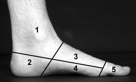
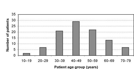
Results: In total, 101 patients were identified. Average age was 47.3 years (range, 14-79 years); there was a marked female preponderance with 73 females and 28 males. Thirty different histological types were identified; ganglion cysts were the most commonly encountered lesions and there was only one malignant lesion encountered in this study. Only 58 out of the 101 lumps were correctly diagnosed prior to surgery. Certain lesions were more commonly encountered in specific zones of the foot.
Conclusions: We have shown that there are a wide variety of potential diagnoses which have to be considered when examining a patient with a foot lump. There is a low diagnostic accuracy for foot lumps and, therefore, surgical excision and histological diagnosis should be sought if there is any uncertainty.
Figures
References
-
- Enzinger FM, Weis SW. Soft tissue tumours. St Louis, MO: Mosby; 1983.
-
- Kirby EJ, Shereff MJ, Lewis MM. Soft-tissue tumors and tumor-like lesions of the foot. An analysis of eighty-three cases. J Bone Joint Surg Am. 1989;71:621–6. - PubMed
-
- Rozbrch SR, Chang V, Bohne WH, Deland JT. Ganglion cysts of the lower extremity: an analysis of 54 cases and review of the literature. Orthopaedics. 1998;21:141–8. - PubMed
-
- Alvi F, Rafee A, Khan T. Tumours on the sole of the foot: a case series. J Bone Joint Surg Br. 2005;87(Suppl 1):80.
-
- Scully SP, Temple HT, Harrelson JM. Synovial sarcoma of the foot and ankle. Clin Orthop. 1999;364:220–6. - PubMed
MeSH terms
LinkOut - more resources
Full Text Sources
Medical