

Accuracy of measurements of mandibular anatomy in cone beam computed tomography images
- PMID: 17395068
- PMCID: PMC3644804
- DOI: 10.1016/j.tripleo.2006.04.008
Accuracy of measurements of mandibular anatomy in cone beam computed tomography images
Abstract
Objectives: Cone beam computed tomography (CBCT) images of ideally positioned and systematically mispositioned dry skulls were measured using two-dimensional and three-dimensional software measurement techniques. Image measurements were compared with caliper measurements of the skulls.
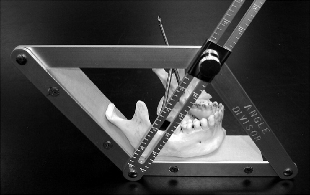
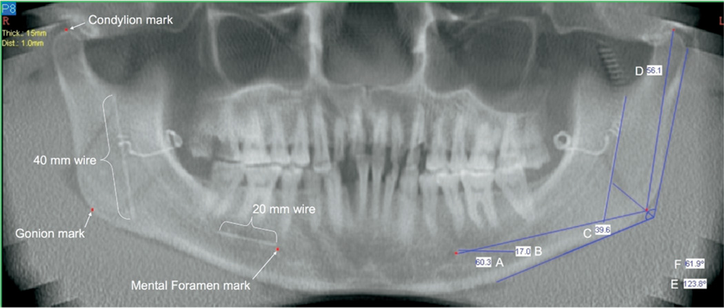
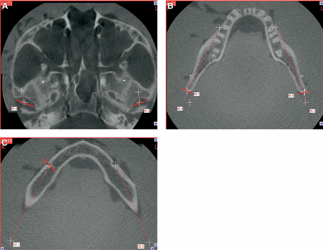
Study design: Cone beam computed tomography volumes of 28 skulls in ideal, shifted, and rotated positions were assessed by measuring distances between anatomic points and reference wires by using panoramic reconstructions (two-dimensional) and direct measurements from axial slices (three-dimensional). Differences between caliper measurements on skulls and software measurements in images were assessed with paired t tests and analysis of variance (ANOVA).
Results: Accuracy of measurement was not significantly affected by alterations in skull position or measurement of right or left sides. For easily visualized orthodontic wires, measurement accuracy was expressed by average errors less than 1.2% for two-dimensional measurement techniques and less than 0.6% for three-dimensional measurement techniques. Anatomic measurements were significantly more variable regardless of measurement technique.
Conclusions: Both two-dimensional and three-dimensional techniques provide acceptably accurate measurement of mandibular anatomy. Cone beam computed tomography measurement was not significantly influenced by variation in skull orientation during image acquisition.
Figures
References
-
- Laster WS, Ludlow JB, Bailey LJ, Hershey HG. Accuracy of measurements of mandibular anatomy and prediction of asymmetry in panoramic radiographic images. Dentomaxillofac Radiol. 2005;34:343–349. - PubMed
-
- Tronje G, Eliasson S, Julin P, Welander U. Image distortion in rotational panoramic radiography. II. Vertical distances. Acta Radiol Diagn (Stockh) 1981;22:449–455. - PubMed
-
- Habets LL, Bezuur JN, van Ooij CP, Hansson TL. The orthopantomogram, an aid in diagnosis of temporomandibular joint problems. I. The factor of vertical magnification. J Oral Rehabil. 1987;14:475–480. - PubMed
-
- Kjellberg H, Ekestubbe A, Kiliaridis S, Thilander B. Condylar height on panoramic radiographs. A methodologic study with a clinical application. Acta Odontol Scand. 1994;52:43–50. - PubMed
-
- Turp JC, Vach W, Harbich K, Alt KW, Strub JR. Determining mandibular condyle and ramus height with the help of an Orthopantomogram–a valid method? J Oral Rehabil. 1996;23:395–400. - PubMed
Publication types
MeSH terms
Grants and funding
LinkOut - more resources
Full Text Sources
