Review
. 2007 May 29;362(1481):877-86.
doi: 10.1098/rstb.2007.2094.

Neural correlates of the contents of visual awareness in humans

Affiliations
Review

Neural correlates of the contents of visual awareness in humans

Geraint Rees. Philos Trans R Soc Lond B Biol Sci. .

Abstract

The immediacy and directness of our subjective visual experience belies the complexity of the neural mechanisms involved, which remain incompletely understood. This review focuses on how the subjective contents of human visual awareness are encoded in neural activity. Empirical evidence to date suggests that no single brain area is both necessary and sufficient for consciousness. Instead, necessary and sufficient conditions appear to involve both activation of a distributed representation of the visual scene in primary visual cortex and ventral visual areas, plus parietal and frontal activity. The key empirical focus is now on characterizing qualitative differences in the type of neural activity in these areas underlying conscious and unconscious processing. To this end, recent progress in developing novel approaches to accurately decoding the contents of consciousness from brief samples of neural activity show great promise.
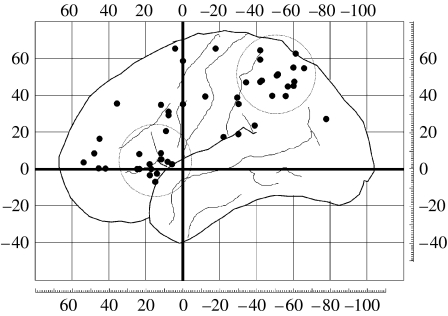

Figures

Figure 1
Figure 1
Unconscious and conscious processing in V1. (a) An oriented target annulus can be effectively rendered invisible by subsequently presenting a contrast-inverted mask with no specific orientation. (b) However, pattern-based decoding applied to V1 activity measured using fMRI can still successfully determine significantly greater than chance which target orientation was presented to a subject. Decoding is not significantly different from chance in V2 and V3. This demonstrates that unconscious information about target orientation must be present in the fMRI signals in V1. Data are from Haynes & Rees (2005a). (c) Apparent motion on a curved path can be perceived by alternating appearance of tilted line inducers on the horizontal or vertical meridians. (d) Perception of apparent motion is associated with significantly enhanced feedback effective connectivity (as assessed with dynamic causal modelling) from V5/MT to the retinotopic location in V1 on the path of apparent motion. Data are from Sterzer et al. (2006).
Figure 2
Figure 2
Unconscious representation of face-specific information in FFA. Prediction accuracy of a multivoxel pattern-based decoder for discriminating the presentation of either a face or house from activity recorded in the fusiform face area (FFA) or parahippocampal place area (PPA), respectively. Average prediction accuracies across participants (n=5) for visible faces versus houses are denoted by filled circles (±s.e.m.) and for invisible faces versus houses by empty circles. Performance was uniformly high and significantly above chance level (50%, dotted line) for all pairwise comparisons across participants.
Figure 3
Figure 3
Decoding the stream of consciousness from activity in human V1. A multivoxel pattern-based decoder trained on activity from V1, while participants experience binocular rivalry can successfully decode the stream of consciousness over several minutes. Data are from Haynes & Rees (2005b) and show participants' reported percepts (dotted lines) alternating between each monocular view. Blind predictions of conscious contents made from patterns of V1 activity by the decoder are shown (solid lines). A close correspondence between decoded and actual perceptual state is apparent.
Figure 4
Figure 4
Fronto-parietal activation associated with awareness. Areas of parietal and prefrontal cortex that show activation correlated with changes in visual awareness (Kleinschmidt et al. 1998; Lumer et al. 1998; Sterzer et al. 2002) are plotted on a template brain. Each circle is placed at the centre of a cluster of activation; overlapping loci from the same study are omitted for clarity. There is prominent clustering of activations in superior parietal and dorsolateral prefrontal cortex, highlighted by large, dotted circles.

References

    1. Barnes J, Howard R.J, Senior C, Brammer M, Bullmore E.T, Simmons A, David A.S. The functional anatomy of the McCollough contingent colour after-effect. Neuroreport. 1999;10:195–199. doi:10.1097/00001756-199901180-00037 - DOI - PubMed
    1. Beck D.M, Rees G, Frith C.D, Lavie N. Neural correlates of change detection and change blindness. Nat. Neurosci. 2001;4:645–650. doi:10.1038/88477 - DOI - PubMed
    1. Beck D.M, Muggleton N, Walsh V, Lavie N. Right parietal cortex plays a critical role in change blindness. Cereb. Cortex. 2006;16:712–717. doi:10.1093/cercor/bhj017 - DOI - PubMed
    1. Bodis-Wollner I, Bucher S.F, Seelos K.C. Cortical activation patterns during voluntary blinks and voluntary saccades. Neurology. 1999;53:1800–1805. - PubMed
    1. Bristow D, Haynes J.D, Sylvester R, Frith C.D, Rees G. Blinking suppresses the neural response to unchanging retinal stimulation. Curr. Biol. 2005;15:1296–1300. doi:10.1016/j.cub.2005.06.025 - DOI - PubMed

Publication types

LinkOut - more resources