Comparative Study
. 2007 Jun 15;581(Pt 3):1001-18.
doi: 10.1113/jphysiol.2007.132886. Epub 2007 Mar 29.

Rat alpha6beta2delta GABAA receptors exhibit two distinct and separable agonist affinities

Affiliations
Comparative Study

Rat alpha6beta2delta GABAA receptors exhibit two distinct and separable agonist affinities

Stephen H Hadley et al. J Physiol. .

Abstract

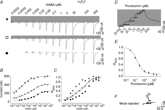
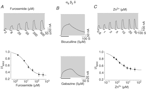
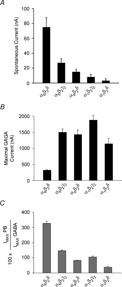
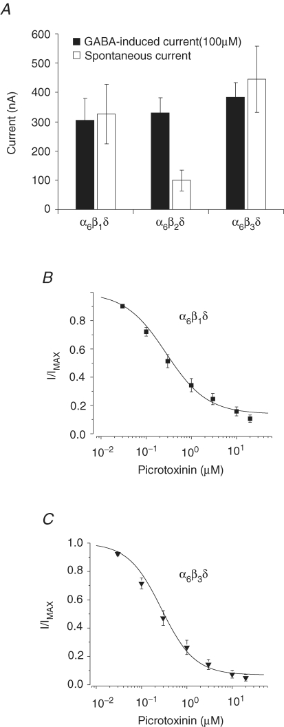
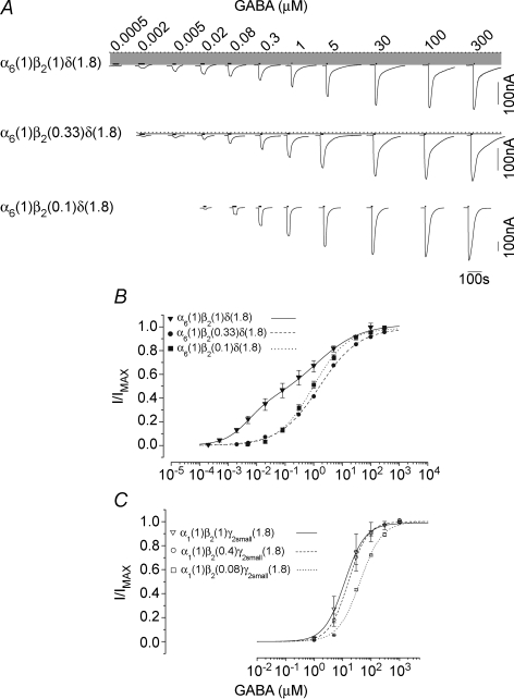
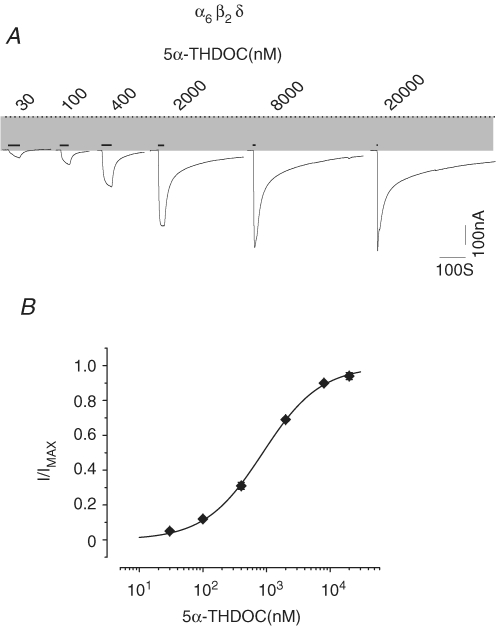
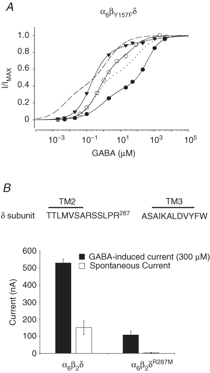
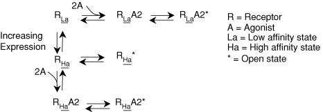
The onset of motor learning in rats coincides with exclusive expression of GABAA receptors containing alpha6 and delta subunits in the granule neurons of the cerebellum. This development temporally correlates with the presence of a spontaneously active chloride current through alpha6-containing GABAA receptors, known as tonic inhibition. Here we report that the coexpression of alpha6, beta2, and delta subunits produced receptor-channels which possessed two distinct and separable states of agonist affinity, one exhibiting micromolar and the other nanomolar affinities for GABA. The high-affinity state was associated with a significant level of spontaneous channel activity. Increasing the level of expression or the ratio of beta2 to alpha6 and delta subunits increased the prevalence of the high-affinity state. Comparative studies of alpha6beta2delta, alpha1beta2delta, alpha6beta2gamma2, alpha1beta2gamma2 and alpha4beta2delta receptors under equivalent levels of expression demonstrated that the significant level of spontaneous channel activity is uniquely attributable to alpha6beta2delta receptors. The pharmacology of spontaneous channel activity arising from alpha6beta2delta receptor expression corresponded to that of tonic inhibition. For example, GABAA receptor antagonists, including furosemide, blocked the spontaneous current. Further, the neuroactive steroid 5alpha-THDOC and classical glycine receptor agonists beta-alanine and taurine directly activated alpha6beta2delta receptors with high potency. Specific mutation within the GABA-dependent activation domain (betaY157F) impaired both low- and high-affinity components of GABA agonist activity in alpha6betaY157Fdelta receptors, but did not attenuate the spontaneous current. In comparison, a mutation located between the second and third transmembrane segments of the delta subunit (deltaR287M) significantly diminished the nanomolar component and the spontaneous activity. The possibility that the high affinity state of the alpha6beta2delta receptor modulates the granule neuron activity as well as potential mechanisms affecting its expression are discussed.


Figures

Figure 1
Figure 1
α6β2δ receptors exhibit spontaneous activity and two separable state of agonist affinities A, representative GABA current traces for high (top), intermediate (middle) and low (bottom) expression conditions with oocytes clamped at −70 mV. The dotted line indicates the zero-current level; the shaded area represents the spontaneous activity. The thick lines above the current traces represent the duration of GABA application. The duration of agonist application decreased with increasing concentration of agonist since the currents reached steady state (peaked) more rapidly at higher concentrations of agonist. B, plot of GABA concentration–response relationships including the spontaneous currents. All data were fitted with a single Hill equation. C, concentration–response relationship of the normalized GABA currents. The EC50, nH and maxima parameters for the group data for high, intermediate and low expression conditions are shown in Table 1. The continuous line shows the plot of the fit of a sum of two Hill equations to the data points from an oocyte with intermediate expression level demonstrating the presence of two components with different sensitivities to GABA. The dashed line shows the overall plot of the fit (group data) of sum of two Hill equations to the GABA concentration–response data points for the α6β2δ receptor at median expression. D, a representative current trace for consecutive application of 0.03–20 μm picrotoxinin on an oocyte with high expression of α6β2δ recptor. The thick lines below the current traces represent the duration of antagonist application. The arrow indicates the start of the picrotoxinin application at the concentration shown below it. E, the concentration–response relationship for picrotoxinin block of the spontaneous current form α6β2δ receptor. Picrotoxinin inhibited greater than 90% of the spontaneous current with an IC50 of approximately 0.2 μm.F, the effect of 20 μm picrotoxinin application on the leak current (20 nA) from a mock-injected oocyte (see methods).
Figure 2
Figure 2
Furosemide, bicuculline, gabazine and Zn2+ inhibit the spontaneous current arising from α6β2δ receptors A, furosemide blocked the spontaneous activity of α6β2δ receptors. Current traces and the concentration–response relationship for furosemide-dependent inhibition of the spontaneous current arising from an oocyte with a high level of expression of α6β2δ receptors. The dotted line indicates the zero-current level; the shaded area represents the spontaneous activity. The thick lines below the current traces represent the duration of antagonist application. B, bicuculline and gabazine inhibited the spontaneous activity of α6β2δ receptors. Current traces representing bicuculline (5 μm) and gabazine (5 μm) inhibitory action on the spontaneous activity. C, the representative current traces and concentration–response relationship for Zn2+ block of the spontaneous current arising from α6β2δ receptors.
Figure 3
Figure 3
Comparison of the spontaneous currents, GABA-induced maxima and pentobarbital relative maxima to GABA for α6β2δ, α6β2γ, α4β2δ, α1β2γ, and α1β2δ receptors under equivalent expression conditions A, comparison of the spontaneous currents for α6β2δ, α6β2γ2, α4β2δ, α1β2γ2 and α1β2δ receptors under equivalent expression conditions. The α6β2δ receptor exhibited a significantly higher spontaneous current than did the other GABAA receptors. B, comparison of the GABA-induced maximal current for α6β2δ, α6β2γ2, α4β2δ, α1β2γ2 and α1β2δ receptors under equivalent expression conditions. GABA maximal current was determined from experiments in A using GABA concentrations 20–50 times the respective EC50 value. GABA had the lowest efficacy (maximal) for α6β2δ receptors. C, comparison of the relative maximal current of pentobarbital to GABA for the GABAA receptor subtypes. Pentobarbital maximal current was determined from experiments in A using 1 mm concentration. Pentobarbital exhibited a significantly higher efficacy than did GABA for α6β2δ receptors.
Figure 4
Figure 4
Expression of the high-affinity state of α6β2δ receptors is independent of the isoform of the β subunit GABA induced similar maximal current for α6β1δ, α6β2δ, and α6β3δ receptors (filled bars) with high levels of spontaneous activity indicating the presence of the high-affinity state (open bars). B and C, the concentration–response relationship for picrotoxinin block of the spontaneous current from α6β1δ and α6β3δ receptors.
Figure 5
Figure 5
Comparison of the I4AA and TACA maximal-induced currents as well as β-alanine and taurine concentration-response relationship for α6β2δ receptors A, comparison of the I4AA and TACA maximal-induced currents relative to GABA for α6β2δ receptors. B, concentration–response relationship for I4AA and the fit of a sum of two Hill equations to these data points. The α6β2δ receptors response to I4AA concentrations exhibited two components with marked difference in apparent affinity. C, the β-alanine concentration–response relationships for oocytes with high (▾), intermediate (○) and low (•) α6β2δ expression. The dashed line shows the plot of the fit (average from group data) of sum of two Hill equations to the β-alanine concentration–response data point for wild-type α6β2δ receptor at intermediate expression condition. D, taurine concentration–response relationships for α6β2δ receptors at low (•), intermediate (○), or high (▾) expression condition. The dashed line shows the plot of the fit (average from group data) using a sum of two Hill equations to the taurine data points for the α6β2δ receptor at intermediate expression condition.
Figure 6
Figure 6
The effect of change in β2 subunit ratio on the expression of α6β2δ and α1β2γ2 receptors A, the current traces of receptor–channels from the expression of 1α6 : 0.1 β2 : 1.8δ, 1α6 : 0.3 β2 : 1.8δ, or control 1α6 : 1 β2 : 1.8δ ratios. The dotted line indicates the zero-current level; the shaded area represents the spontaneous activity. The thick lines above the current traces represent the duration of GABA application. B, the GABA concentration–response relationship for the three tested ratios of α6, β2, and δ subunits. The data derived from 1α6 : 0.1 β2 : 1.8δ and 1α6 : 0.3 β2 : 1.8δ were fitted with a single Hill equation, while the data for 1α6 : 1 β2 : 1.8δ were fitted with a sum of two Hill equations. C, the concentration–response relationship for 1α1 : 0.08 β2 : 1.8γ2S, 1α1 : 0.4 β2 : 1.8γ2S and control1α1 : 1 β2 : 1.8γ2S subunit combination.
Figure 7
Figure 7
The 5α-THDOC directly activates α6β2δ receptors A, 5α-THDOC-induced current traces. The dotted line indicates the zero-current level; the shaded area represents the spontaneous activity. The thick lines above the current traces represent the duration of 5α-THDOC applications. B, the 5α-THDOC concentration–response relationship and fit of data point to a Hill equation.
Figure 8
Figure 8
The effect of β2 or δ subunit mutation on the expression of α6β2δ receptors A, the GABA concentration–response relationship for α6βY157Fδ receptors for three oocytes with 30 nA (•), 70 nA (○) and 180 nA (▾) of spontaneous current. The mutation within the GABA-dependent activation domain (α6βY157Fδ) did not abolish the spontaneous activity. The continuous lines represent the fit of the GABA data points with a sum of two Hill equations for α6βY157Fδ receptors at different expression levels. The dashed line shows the overall plot of the fit of the GABA concentration–response relationship data with a sum of two Hill equations for the wild-type α6β2δ receptor (intermediate expression). The dotted line shows a plot of the fit (group data) of the GABA concentration–response relationship data with a sum of two Hill equations for the α6βY157Fδ receptor. B, the δR287M mutation attenuated the spontaneous activity and the GABA maximal current. Top shows the amino acid sequence between the TM2 and TM3 domains of the δ subunit, indicating the position of Arg287. At equivalent expression conditions, the wild-type receptor showed high levels in comparison to no spontaneous activity for α6β2δR287M receptors (corrected for control leak, see methods). The maximal-induced GABA current for mutated receptor was significantly reduced as compared to that for wild-type receptor.
Figure 9
Figure 9
A model showing different states of α6β2δ receptors upon clustering

Similar articles

Cited by

References

    1. Amin J. A single hydrophobic residue confers barbiturate sensitivity to γ-aminobutyric acid type C receptor. Mol Pharmacol. 1999;55:411–423. - PubMed
    1. Amin J, Weiss DS. GABAA receptor needs two homologous domains of the β-subunit for activation by GABA but not by pentobarbital [see comment] Nature. 1993;366:565–569. - PubMed
    1. Amin J, Weiss DS. Insights into the activation mechanism of rho1 GABA receptors obtained by coexpression of wild type and activation-impaired subunits. Proc Biol Sci. 1996;263:273–282. - PubMed
    1. Baulac S, Huberfeld G, Gourfinkel-An I, Mitropoulou G, Beranger A, Prud'homme JF, Baulac M, Brice A, Bruzzone R, LeGuern E. First genetic evidence of GABAA receptor dysfunction in epilepsy: a mutation in the γ2-subunit gene. Nat Genet. 2001;28:46–48. - PubMed
    1. Baumann SW, Baur R, Sigel E. Subunit arrangement of γ-aminobutyric acid type A receptors. J Biol Chem. 2001;276:36275–36280. - PubMed

Publication types

MeSH terms

Substances

LinkOut - more resources