. 2007 Aug 1;582(Pt 3):927-37.
doi: 10.1113/jphysiol.2007.132795. Epub 2007 Mar 29.

Imaging and manipulating phosphoinositides in living cells

Affiliations

Imaging and manipulating phosphoinositides in living cells

Tamas Balla. J Physiol. .

Abstract

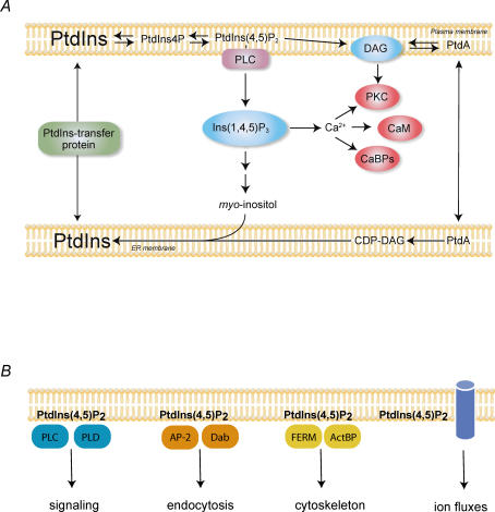
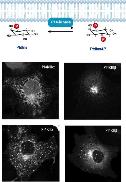
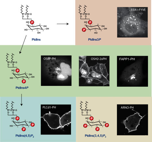
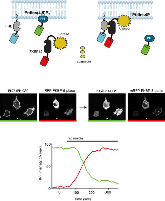
Phosphoinositides are minor phospholipid constituents of virtually every biological membrane yet they play fundamental roles in controlling membrane-bound signalling events. Phosphoinositides are produced from phosphatidylinositol (PtdIns) by phosphorylation of one or more of three positions (3, 4 and 5) of the inositol headgroup located at the membrane cytoplasmic interface by distinct families of inositol lipid kinases. Intriguingly, many of the kinase reactions are catalysed by more than one form of the kinases even in simple organisms and these enzymes often assume non-redundant functions. A similar diversity is seen with inositide phosphatases, the enzymes that dephosphorylate phosphoinositides with a certain degree of specificity and the impairments of which are often linked to human diseases. This degree of multiplicity at the enzyme level together with the universal roles of these lipids in cell regulation assumes that inositol lipids are spatially and functionally restricted in specific membrane compartments. Studying the compartmentalized roles of these lipids at the cellular level represents a major methodological challenge. Over the last 10 years significant progress has been made in creating reagents that can monitor inositol lipid changes in live cells with fluorescence or confocal microscopy. New methods are also being developed to manipulate these lipids in specific membrane compartments in a regulated fashion. This article recalls some historical aspects of inositide research and describes the new methodological advances highlighting their great potential as well as the problems one can encounter with their use.


Figures

Figure 1
Figure 1. The phosphoinositide signalling cascade and processes at the PM controlled by PtdIns(4,5)P2
A, the canonical phosphoinositide signalling cascade. PM PtdIns(4,5)P2 is hydrolysed by activated PLC enzymes upon receptor stimulation to yield the two messengers, Ins(1,4,5)P3 and DAG. Ins(1,4,5)P3 mobilizes intracellular Ca2+ via its receptors located in the ER that function as Ca2+ channels. DAG helps recruit PKC enzymes to the membrane and the cytosoloic Ca2+ increase stimulates Ca2+ sensitive enzymes such as calmodulin (CaM) or other Ca2+ binding proteins (CaBPs) to regulate downstream effectors. Ins(1,4,5)P3 is rapidly degraded by sequential dephosphorylations to myo-inositol, which is then reused for PtdIns synthesis in the ER by conjugation with CDP-DAG. PtdIns transfer proteins facilitate the movement of PtdIns between membranes. B, PtdIns(4,5)P2 controls numerous processes at the PM. It activates enzymes such as PLCδ and PLD, it helps recruit clathrin adaptor proteins, such as AP-2 or Dab2, it provides a link between the membrane and the cytoskeleton via interaction with the FERM domain containing or other actin binding proteins (ActBP) and also regulates ion channels and transporters. It is very likely that the affinities of interaction of the lipid with the various proteins show big variations and when the overall level of the lipid drops in the membrane, it will affect processes that are controlled by weaker interactions. In contrast, effectors with tight PtdIns(4,5)P2 binding are less affected by global PtdIns(4,5)P2 changes but perhaps are sensitive to very local changes that are evoked by colocalized kinases or phosphatases.
Figure 2
Figure 2. Inositide kinase reactions are often performed by multiple enzymes
Phosphorylation of PtdIns to PtdIns4P is mediated by four distinct PI 4-kinases (PI4Ks) that localize to unique membrane compartments. All of the enzymes show localization to the Golgi compartment but PI4KIIIα is also found in the ER, while PI4KIIα is also found in endosomes and the PM. PI4IIIβ is exclusively localized to the Golgi while PI4KIIβ shows partial Golgi and some endosomal localization. The localization of the enzymes does not show dramatic changes after stimulation of the cells even when PtdIns4P levels are expected to change in compartments where the enzymes are not enriched (e.g. the PM). This indicates that there is a need to detect their lipid product, PtdIns4P, in addition to following the enzyme distribution to better understand their regulation.
Figure 3
Figure 3. Steady-state distribution of inositide detecting probes visualized in live cells
The structure of the inositide headgroup is shown along with the cellular distribution of the particular protein module recognizing the lipid and fused to the enhanced green fluorescent protein (GFP). Note that PtdIns4P can be detected either in the PM or in the Golgi, depending on the probe used. No such discrepancy has been found so far with probes detecting PtdIns(4,5)P2, PtdIns3P and PtdIns(3,4,5)P2 but it is still not certain that this means that these lipids are only found in those membranes. More probes are needed to investigate this question.
Figure 4
Figure 4. Chemically induced manipulation of PtdIns(4,5)P2 in the PM
The FRB fragment of the mTOR protein is targeted to the PM by an N-terminal palmitoylation sequence and is also tagged with CFP (this channel is not shown). The 5-phosphatase (5-ptase) domain of the type-IV phosphoinositide 5-phosphatase enzyme is fused to an mRFP–FKBP12 construct and this protein is cytosolic under basal conditions (see image on the left with red bar). Simultaneous expression of the PH domain of PLCδ1 fused to GFP shows PtdIns(4,5)P2 in the PM (image on the left with green bar). Addition of rapamycin heterodimerizes the FRB–FKBP12 pairs and thereby recruits the 5-phosphatase to the membrane (image on the right with red bar) which, in turn, rapidly eliminates PtdIns(4,5)P2 as indicated by the translocation of PLCδ1PH–GFP from the membrane to the cytosol (image on the right with green bar). These changes can be monitored by TIRF microscopy as indicated by the bottom graph. The fluorescence intensity of the PLCδ1PH–GFP in the membrane plane drops rapidly from its initial high value after rapamycin addition (green trace) as the 5-phosphatase is recruited to the PM (red trace).

References

    1. Astle MV, Seaton G, Davies EM, Fedele CG, Rahman P, Arsala L, Mitchell CA. Regulation of phosphoinositide signaling by the inositol polyphosphate 5-phosphatases. IUBMB Life. 2006;58:451–456. - PubMed
    1. Attree O, Olivos IM, Okabe I, Bauiley LC, Nelson DL, Lewis RA, Mcinnes RR, Nussbaum RL. The Lowe's oculocerebrorenal syndrome gene encodes a protein highly homologous to inositol polyphosphate-5-phosphatase. Nature. 1992;358:239–242. - PubMed
    1. Balla A, Balla T. Phosphatidylinositol 4-kinases; old enzymes with emerging functions. Trends Cell Biol. 2006;16:351–361. - PubMed
    1. Balla A, Tuymetova G, Tsiomenko A, Varnai P, Balla T. A plasma membrane pool of phosphatidylinositol 4-phosphate is generated by phosphatidylinositol 4-kinase type-III α: studies with the PH domains of the oxysterol binding protein and FAPP1. Mol Biol Cell. 2005;16:1282–1295. - PMC - PubMed
    1. Ballou CE, Lee YC. The structure of a myoinositol mannoside from Myobacterium tuberculosis glycolipid. Biochemistry. 1964;3:682–685. - PubMed

Publication types

MeSH terms

LinkOut - more resources