. 2007 Apr;56(4):960-7.
doi: 10.2337/db06-1254.

Ischemic vascular damage can be repaired by healthy, but not diabetic, endothelial progenitor cells

Affiliations

Ischemic vascular damage can be repaired by healthy, but not diabetic, endothelial progenitor cells

Sergio Caballero et al. Diabetes. 2007 Apr.

Abstract

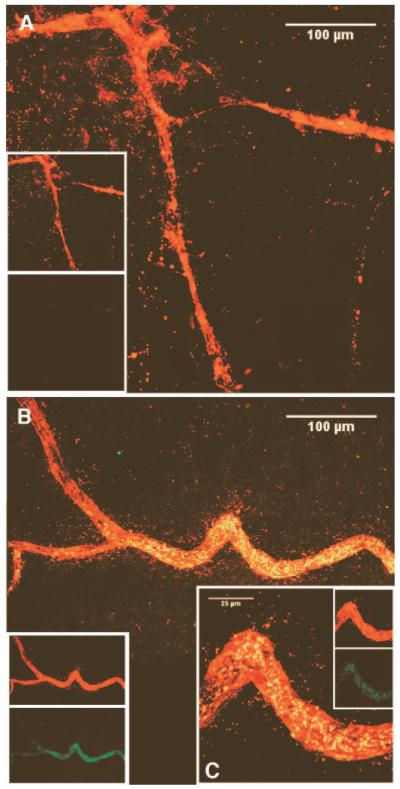
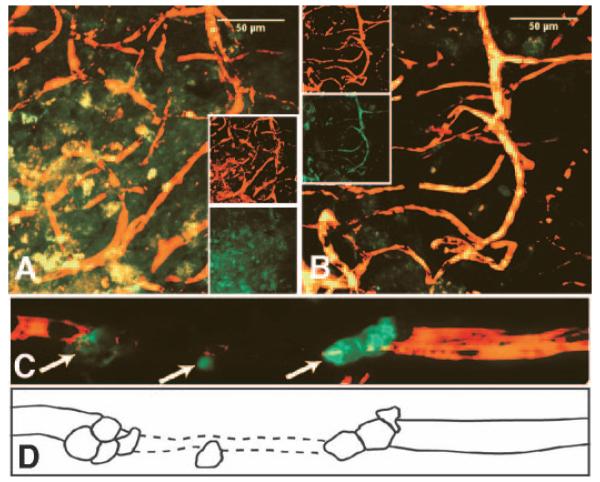
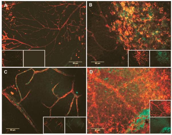
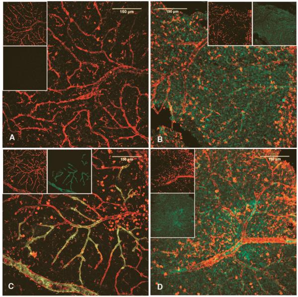
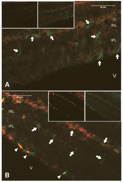
Endothelial precursor cells (EPCs) play a key role in vascular repair and maintenance, and their function is impeded in diabetes. We previously demonstrated that EPCs isolated from diabetic patients have a profound inability to migrate in vitro. We asked whether EPCs from normal individuals are better able to repopulate degenerate (acellular) retinal capillaries in chronic (diabetes) and acute (ischemia/reperfusion [I/R] injury and neonatal oxygen-induced retinopathy [OIR]) animal models of ocular vascular damage. Streptozotocin-induced diabetic mice, spontaneously diabetic BBZDR/Wor rats, adult mice with I/R injury, or neonatal mice with OIR were injected within the vitreous or the systemic circulation with fluorescently labeled CD34(+) cells from either diabetic patients or age- and sex-matched healthy control subjects. At specific times after administering the cells, the degree of vascular repair of the acellular capillaries was evaluated immunohistologically and quantitated. In all four models, healthy human (hu)CD34(+) cells attached and assimilated into vasculature, whereas cells from diabetic donors uniformly were unable to integrate into damaged vasculature. These studies demonstrate that healthy huCD34(+) cells can effectively repair injured retina and that there is defective repair of vasculature in patients with diabetes. Defective EPCs may be amenable to pharmacological manipulation and restoration of the cells' natural robust reparative function.


Figures

FIG. 1
FIG. 1
Injection of huCD34+ cells within the vitreous results inreendothelialization of damaged vessels in the mouse model of OIR. Green-labeled huCD34+ cells were administered to mouse pups (n = 8) on P12 and eyes harvested at P14. Images are z-series projections of two-color LSCM. Panel A depicts the central region of a retina from a mouse that did not receive huEPC, and shows expected pathology, including lack of microvessels and degenerate larger vessels. Panel B depicts a similar region of a retina from a mouse eye that was injected with huEPC. Note the high degree of incorporation of labeled cells into the dilated and functioning vessel (yellow). Inset panel C is a higher magnification of a portion of the vessel in panel B. The other insets show separate red and green channels used to make the composite images.
FIG. 2
FIG. 2
huEPC of nondiabetic, but not diabetic, origin integrate into degenerate vasculature in mouse eyes damaged by I/R injury (n = 20). huCD34+ cells were given by intravitreous injection (n = 10) or systemically (n = 10) 7 days after I/R injury. Two days later, animals were killed and perfused with rhodamine-conjugated dextran to examine vessel patency. Cells of diabetic origin (panel A) do not integrate into existing vasculature, whereas cells of nondiabetic origin (panel B) show extensive integration into small and medium sized vessels (yellow in the composite images). Images are z-series projections of two-color LSCM. Insets show separate red and green channels used to make the composite images. In panel C CD34+ cells home to an area of injury and traverse toward the ischemic region of the capillary (arrows). Panel D is a schematic representation of the image in panel C and depicts the nonperfused/acellular region as a hatched line in order to better visualize the process.
FIG. 3
FIG. 3
Normal huEPC, but not diabetic huEPC, participate in ocular vascular reendothelialization in STZ-diabetic mice. All images are composite red and green channels (shown in respective insets) of epifluorescence digital captures of representative neural retinas. STZ-diabetic (n = 12) and age- and sex-matched normal control mice (n = 12) were given labeled huCD34+ cells from either diabetic (n = 6 diabetic mice, n = 6 nondiabetic mice) or nondiabetic donors (n = 6 diabetic mice, n = 6 nondiabetic mice), and eyes were harvested 48 h later. Panel A is from a normal mouse that received nondiabetic cells. Lack of vascular injury should preclude incorporation of labeled EPC. Note the lack of labeled cells. Panel B is from a normal mouse that received diabetic huEPC. Note the tendency for these labeled cells to form clumps distinct from the vasculature. By contrast, panel C shows extensive incorporation of labeled CD34+ cells from a normal donor in damaged vasculature of a STZ-diabetic mouse. Panel D demonstrates that huEPC incorporation is not solely a function of vascular damage in the recipient, since labeled diabetic CD34+ cells do not integrate into vasculature of a STZ-diabetic mouse, but rather form sheets and clumps separate from the vascular plexus.
FIG. 4
FIG. 4
huCD34+ EPC of nondiabetic, but not diabetic, origin integrate into damaged vasculature in BBZ/Wor diabetic rats. Labeled cells were given by intravitreous injection to immune suppressed rats (n = 6 nondiabetic rats/nondiabetic EPC; n = 6 nondiabetic rats/diabetic EPC; n = 6 diabetic rats/nondiabetic EPC; n = 6 diabetic rats/diabetic EPC). All images are z-series projections of two-color LSCM, with insets showing respective separate red and green channels. Panel A is from a healthy rat receiving nondiabetic huEPC. Note the lack of labeled cells consistent with lack of vascular injury. Panel B shows that diabetic huEPC in a normal rat eye form a discrete layer without integrating into vessels. Panel C demonstrates clear reendothelialization by labeled normal huEPC into presumably damaged vasculature of a diabetic rat. Panel D shows again that diabetic EPC are incapable of integrating into damaged vasculature, instead forming distinct clumps and layers separate from vessels.
FIG. 5
FIG. 5
EPC integrate into damaged vessel walls and are not merely associated perivascularly with vessels. Two-color fluorescent immunohistology of frozen sections of mouse eyes with intravitreous huEPC shows green mouse endothelial cells (arrows) in the superficial, middle, and deep vascular plexi of the neural retina of both normal control (n = 4) (panel A) and STZ-diabetic (n = 4) (panel B) mice. However, only in the diabetic mouse eye in panel B are there red huEPC (arrowheads), which align with the mouse endothelium. V = vitreous; INL = inner nuclear layer; IPL = inner plexiform layer.

References

    1. Fadini GP, Sartore S, Baesso I, Lenzi M, Agostini C, Tiengo A, Avogaro A. Endothelial progenitor cells and the diabetic paradox. Diabetes Care. 2006;29:714–716. - PubMed
    1. Awad O, Dedkov E, Jiao C, Bloomer S, Tomanek RJ, Schatteman GC. Differential healing activities of CD34+ and CD14+ endothelial cell progenitors. Arterioscler Thromb Vasc Biol. 2006;26:758–764. - PubMed
    1. Asahara T, Masuda H, Takahashi T, Kalka C, Pastore C, Silver M, Kearne M, Magner M, Isner JM. Bone marrow origin of endothelial progenitor cells responsible for postnatal vasculogenesis in physiological and pathological neovascularization. Circ Res. 1999;85:221–228. - PubMed
    1. Asahara T, Murohara T, Sullivan A, Silver M, van der Zee R, Li T. Isolation of putative progenitor endothelial cells for angiogenesis. Science. 1997;275:964–967. - PubMed
    1. Grant MB, May WS, Caballero S, Brown GA, Guthrie SM, Mames RN, Byrne BJ, Vaught T, Spoerri PE, Peck AB, Scott EW. Adult hematopoietic stem cells provide functional hemangioblast activity during retinal neovascularization. Nat Med. 2002;8:607–612. - PubMed

Publication types