. 2007 Jun;292(6):G1770-83.
doi: 10.1152/ajpgi.00249.2006. Epub 2007 Mar 29.

Enteroendocrine cells express functional Toll-like receptors

Affiliations

Enteroendocrine cells express functional Toll-like receptors

Milena Bogunovic et al. Am J Physiol Gastrointest Liver Physiol. 2007 Jun.

Abstract

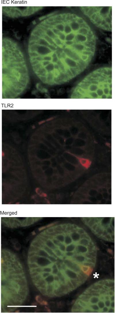
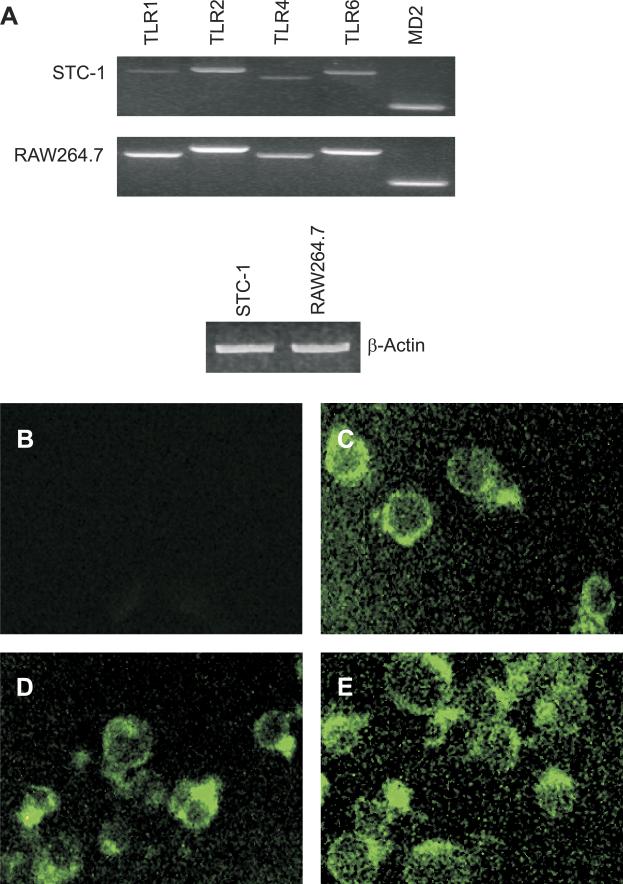
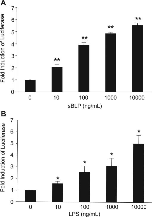
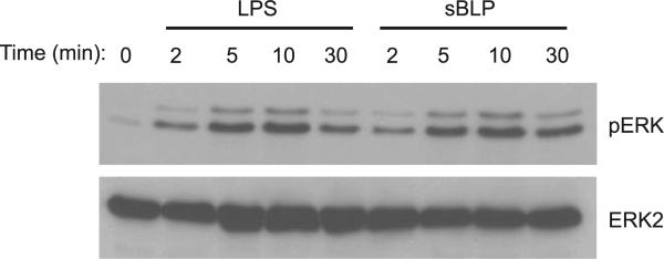
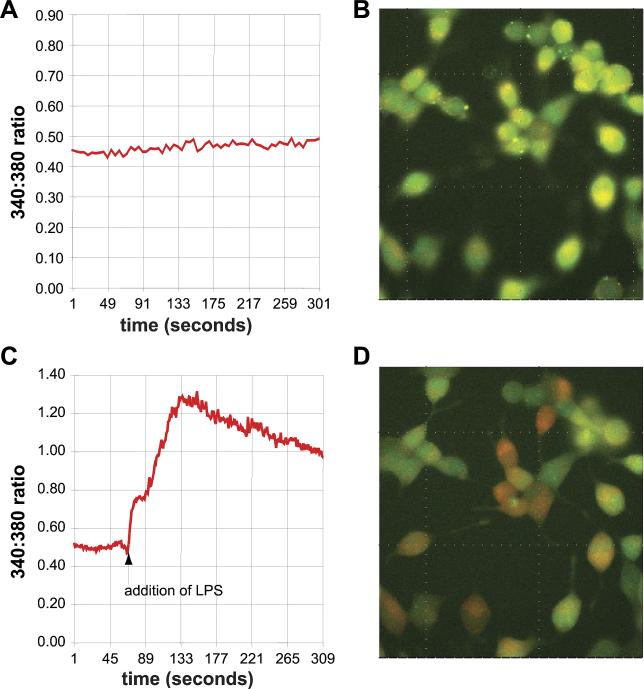
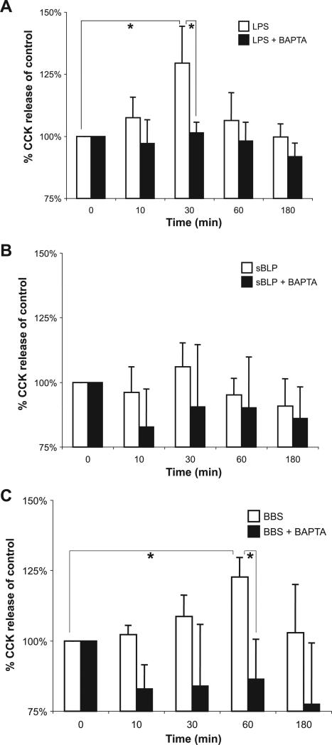
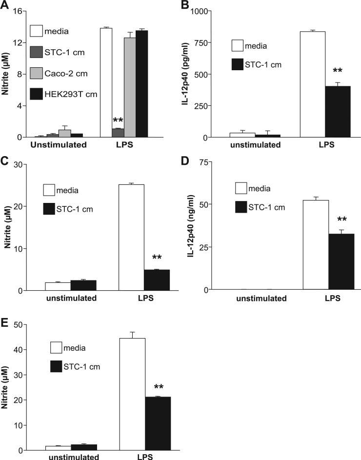
Intestinal epithelial cells (IECs) provide a physical and immunological barrier against enteric microbial flora. Toll-like receptors (TLRs), through interactions with conserved microbial patterns, activate inflammatory gene expression in cells of the innate immune system. Previous studies of the expression and function of TLRs in IECs have reported varying results. Therefore, TLR expression was characterized in human and murine intestinal sections, and TLR function was tested in an IEC line. TLR1, TLR2, and TLR4 are coexpressed on a subpopulation of human and murine IECs that reside predominantly in the intestinal crypt and belong to the enteroendocrine lineage. An enteroendocrine cell (EEC) line demonstrated a similar expression pattern of TLRs as primary cells. The murine EEC line STC-1 was activated with specific TLR ligands: LPS or synthetic bacterial lipoprotein. In STC-1 cells stimulated with bacterial ligands, NF-kappaB and MAPK activation was demonstrated. Furthermore, the expression of TNF and macrophage inhibitory protein-2 were induced. Additionally, bacterial ligands induced the expression of the anti-inflammatory gene transforming growth factor-beta. LPS triggered a calcium flux in STC-1 cells, resulting in a rapid increase in CCK secretion. Finally, conditioned media from STC-1 cells inhibited the production of nitric oxide and IL-12 p40 by activated macrophages. In conclusion, human and murine IECs that express TLRs belong to the enteroendocrine lineage. Using a murine EEC model, a broad range of functional effects of TLR activation was demonstrated. This study suggests a potential role for EECs in innate immune responses.


Figures

Fig. 1
Fig. 1
Toll-like receptor (TLR)1, TLR2, and TLR4 were coexpressed by a human intestinal epithelial cell (IEC) that mainly resides in the crypts. Paraffin-embedded tissue sections of the histologically normal human colon and ileum were immunostained with specific antibodies as described in detail in MATERIALS AND METHODS (A–C). A: double-immunofluorescent staining of the colon with anti-TLR1 (FITC) and anti-TLR2 (Texas red) antibodies. Asterisks show TLR1+TLR2+ cells in the epithelial monolayer. B: double-immunofluorescent staining of the colon with anti-TLR4 (FITC) and anti-TLR1 (Texas red) antibodies. Asterisks show TLR1+TLR4+ cells in the epithelial monolayer. Magnification: ×400. Scale bars = 50 μm. C: double staining of the colon with anti-TLR1 (Texas red; left) and anti-TLR2 (FITC; right) antibodies and a superimposed image of both (middle). A single epithelial cell located in a crypt was visualized by confocal microscopy.
Fig. 2
Fig. 2
TLR2+ cells in the epithelial monolayer of the human colon express IEC-specific cytokeratins. Double staining of the colon with IEC-specific anti-multicytokeratin (FITC) and anti-TLR2 (Texas red) antibodies is shown. The asterisk shows a TLR2+multicytokeratin+ cell in the intestinal crypt. Magnification: ×400. Scale bar = 50 μm.
Fig. 3
Fig. 3
TLR-immunoreactive cells in human colonic specimens express an enteroendocrine cell (EEC) marker, serotonin. A: double staining of the colon with anti-serotonin (FITC) and anti-TLR1 (Texas red) antibodies. The asterisk shows a TLR1+serotonin+ cell in the intestinal crypt. Magnification: ×400. Scale bar = 50 μm. B: double staining of the colon with anti-serotonin (FITC) and anti-TLR2 (Texas red) antibodies. Asterisks show TLR2+serotonin+ cells in the intestinal crypt. Magnification: ×400. Scale bar = 50 μm. IECs expressing TLRs have features reminiscent of an EEC lineage, including a pyramidal shape with a large basolateral surface, an apically displaced nucleus, and a granular appearance.
Fig. 4
Fig. 4
TLR-immunoreactive cells in murine colonic specimens express an EEC marker, serotonin. A and B: double staining of murine colonic sections with anti-TLR2 (Alexa488 secondary; A) and anti-serotonin (Cy3 secondary; B), an EEC marker. C: superimposed image of A and B with nuclei depicted in white and actin in blue. D and E: double staining of murine colonic sections with anti-TLR6 (Alexa488 secondary; D) and anti-serotonin (Cy3 secondary; E). F: superimposed image of D and E with nuclei depicted in white and actin in blue.
Fig. 5
Fig. 5
The murine EEC line STC-1 expresses TLRs. A, top: expressions of TLR and MD2 mRNA were analyzed by PCR of reverse-transcribed total RNA isolated from STC-1 cells. RNA isolated from RAW264.7 macrophage cells was used as a positive control. RT-PCR amplified β-actin mRNA is shown as a loading control (bottom). B–E: STC-1 cells were stained (FITC secondary) with control IgG (B), anti-TLR2 (C), anti-TLR4/MD2 complex (D), and anti-TLR6 antibodies (E). Experiments were repeated 3 times, and a representative result is displayed.
Fig. 6
Fig. 6
TLR1/2 [synthetic bacterial lipoprotein (sBLP)] and TLR4 (LPS) ligands activate NF-κB in the murine STC-1 EEC line. NF-κB activation was assessed by transient transfection of a plasmid with a multimerized NF-κB DNA binding element luciferase reporter. A and B: STC-1 cells were stimulated for 8 h with a dose titration of sBLP (A) or LPS (B). Results are expressed as relative light units normalized to β-galactosidase activity from a cotransfected heat shock protein (HSP) promoter-β-galactosidase plasmid to correct for transfection efficiency. The fold induction of NF-κB luciferase was compared with values in unstimulated cells (equal to 1) for each group. Each result represents the mean ± SD of 3–5 experiments. *P < 0.05 and **P < 0.005 compared with unstimulated cells for each group.
Fig. 7
Fig. 7
sBLP and LPS activate the ERK MAPK pathway in STC-1 cells. MAPK activation was assessed in cells stimulated with LPS or sBLP (10 μg/ml) for 0, 2, 5, 10, or 30 min. Whole cell extracts were isolated as described in MATERIALS AND METHODS. Western blots were probed for phosphorylated (p)ERK1/2 and total ERK2 to determine equal loading. Experiments were repeated 3 times, and a representative result is displayed.
Fig. 8
Fig. 8
TLR ligands induce TNF and macrophage inhibitory protein (MIP)-2 mRNA and protein expression in STC-1 cells. STC-1 cells were stimulated with 10 μg/ml of LPS or sBLP for 0–4 h. A: cells were harvested via TRIzol. Total RNA was isolated, digested with DNase I, and subsequently reverse transcribed into cDNA. TNF, MIP-2, and β-actin PCR primer sequences are described in MATERIALS AND METHODS. Experiments were repeated 3 times, and a representative result is displayed. B–E: STC-1 cells were stimulated with increasing amounts of LPS (B and D) or sBLP (C and E) for 48 h. ELISA was performed on cell-free supernatants for MIP-2 (B and C) and TNF (D and E). Experiments were performed in triplicate, and results represent means ± SD. *P < 0.05, **P < 0.01, and ***P < 0.001 compared with unstimulated cells for each group.
Fig. 9
Fig. 9
LPS and TLR4 induce transforming growth factor (TGF)-β expression in STC-1 cells. A: STC-1 cells were grown in 1% FBS and stimulated with 10 μg/ml LPS for 0–6 h. Total RNA was isolated and reverse transcribed. TGF-β and β-actin primer sequences are described in MATERIALS AND METHODS. Experiments were repeated 3 times, and a representative result is displayed. B: STC-1 cells were stimulated with 0.1–10 μg/ml LPS for 24, 48, or 72 h. ELISA was performed on the supernatants for TGF-β according to the manufacturer's protocols. Experiments were performed in triplicate, and results represent means ± SD. *P < 0.05 compared with unstimulated cells for each time group. C: constitutively active (ca)-TLR4 expression plasmid increased TGF-β production in transfected STC-1 cells. STC-1 cells were grown in 1% FBS and transfected with a total of 1 μg plasmid DNA (empty vector pcDNA3 + ca-TLR4). STC-1 cells were incubated for 48, 72, or 96 h. ELISA was performed on the supernatants for TGF-β according to the manufacturer's protocols. Experiments were performed in triplicate, and results represent means ± SD. *P < 0.05 compared with empty vector control for each time group.
Fig. 10
Fig. 10
LPS induces a rapid calcium flux in STC-1 cells. STC-1 cells were loaded with fura-2 and incubated in DMEM with 10% FBS. A and B: cells were initially stabilized. C and D: cells were subsequently treated with 1 μg/ml LPS (addition of LPS), causing an increase in the internal calcium concentration. Experiments were performed in trip-licate, and a representative result is shown.
Fig. 11
Fig. 11
LPS induces secretion of CCK in STC-1 cells. Cells were pretreated in the presence or absence of the calcium chelator BAPTA-AM (20 μM) 30 min prior to being stimulated for 10, 30, 60, and 180 min with 1 μg/ml LPS (A), 10 μg/ml sBLP (B), or 10 nM bombesin (BBS; C). Enzyme immunoassay was performed on cell-free supernatants for CCK. Experiments were performed 4 times. Due to the variation of the baseline production of CCK, results were normalized to controls (unstimulated) and presented as percentages of control values. The average (mean + SD) baseline production of CCK was 1.8 ± 0.6 ng/ml. Maximal stimulated CCK expression was 2.0 + 0.5 ng/ml for LPS, 1.8 + 0.6 ng/ml for sBLP, and 2.1 + 0.8 for BBS; n = 4 experiments for each condition. *P < 0.05.
Fig. 12
Fig. 12
STC-1 conditioned media (cm) inhibits LPS-stimulated IL-12 p40 and nitric oxide (NO) production in macrophages. A–E: 4-day conditioned media from STC-1 cells were added to RAW264.7 cells (A), murine peritoneal macrophages (B and C), or murine bone marrow (BM)-derived macrophages (D and E). A: 4-day conditioned media from the IEC line Caco-2 and epithelial cell line HEK-293T were used as controls. Macrophages were subsequently stimulated with 1 μg/ml LPS for 24 h (A, B, and D) or 48 h (C and E). Experiments were performed in trip-licate, and results represent means ± SD. **P < 0.01 compared with media control for each group.
Fig. 13
Fig. 13
TGF-β inhibits LPS-stimulated IL-12 p40 and NO production in macrophages. BM-derived (A) or peritoneal (B) macrophages were incubated with or without 5 ng/ml recombinant human (rh)TGF-β1 prior to being stimulated with 1 μg/ml LPS. Supernatants were assayed for NO (A) and IL-12 p40 (B). Experiments were performed in triplicate, and results represent means ± SD. *P < 0.05 and ***P < 0.001 compared with media control for each group.

References

    1. Abad C, Martinez C, Juarranz MG, Arranz A, Leceta J, Delgado M, Gomariz RP. Therapeutic effects of vasoactive intestinal peptide in the trinitrobenzene sulfonic acid mice model of Crohn's disease. Gastroenterology. 2003;124:961–971. - PubMed
    1. Abreu MT, Vora P, Faure E, Thomas LS, Arnold ET, Arditi M. Decreased expression of Toll-like receptor-4 and MD-2 correlates with intestinal epithelial cell protection against dysregulated proinflammatory gene expression in response to bacterial lipopolysaccharide. J Immunol. 2001;167:1609–1616. - PubMed
    1. Barg S. Mechanisms of exocytosis in insulin-secreting B-cells and glucagon-secreting A-cells. Pharmacol Toxicol. 2003;92:3–13. - PubMed
    1. Beck PL, Rosenberg IM, Xavier RJ, Koh T, Wong JF, Podolsky DK. Transforming growth factor-beta mediates intestinal healing and susceptibility to injury in vitro and in vivo through epithelial cells. Am J Pathol. 2003;162:597–608. - PMC - PubMed
    1. Cario E, Podolsky DK. Differential alteration in intestinal epithelial cell expression of toll-like receptor 3 (TLR3) and TLR4 in inflammatory bowel disease. Infect Immun. 2000;68:7010–7017. - PMC - PubMed

Publication types

MeSH terms

Substances

LinkOut - more resources