. 2007 Apr 18;26(8):2218-26.
doi: 10.1038/sj.emboj.7601664. Epub 2007 Mar 29.

Electron cryotomography of immature HIV-1 virions reveals the structure of the CA and SP1 Gag shells

Affiliations

Electron cryotomography of immature HIV-1 virions reveals the structure of the CA and SP1 Gag shells

Elizabeth R Wright et al. EMBO J. .

Abstract

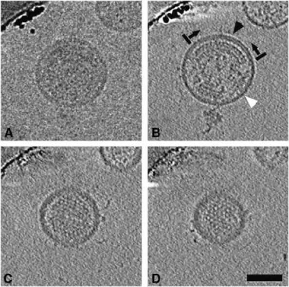
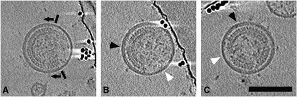
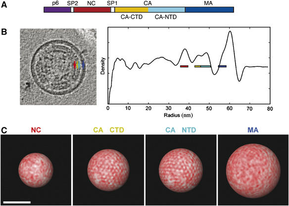
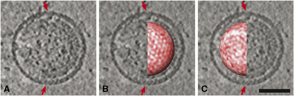
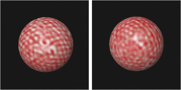
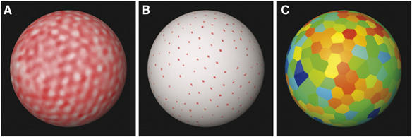
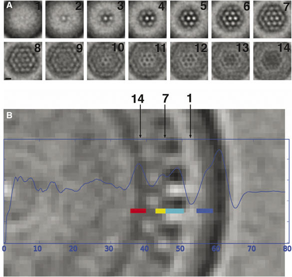
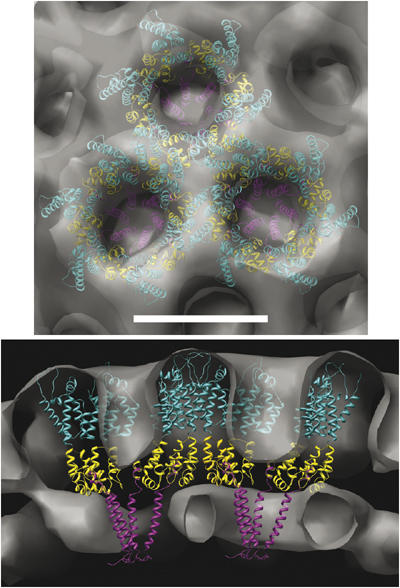
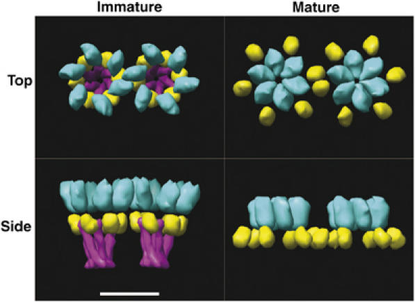
The major structural elements of retroviruses are contained in a single polyprotein, Gag, which in human immunodeficiency virus type 1 (HIV-1) comprises the MA, CA, spacer peptide 1 (SP1), NC, SP2, and p6 polypeptides. In the immature HIV-1 virion, the domains of Gag are arranged radially with the N-terminal MA domain at the membrane and C-terminal NC-SP2-p6 region nearest to the center. Here, we report the three-dimensional structures of individual immature HIV-1 virions, as obtained by electron cryotomography. The concentric shells of the Gag polyprotein are clearly visible, and radial projections of the different Gag layers reveal patches of hexagonal order within the CA and SP1 shells. Averaging well-ordered unit cells leads to a model in which each CA hexamer is stabilized by a bundle of six SP1 helices. This model suggests why the SP1 spacer is essential for assembly of the Gag lattice and how cleavage between SP1 and CA acts as a structural switch controlling maturation.


Figures

Figure 1
Figure 1
Example raw image and reconstruction. (A) The central image from a tilt series of an isolated immature HIV-1 virion suspended in vitreous ice. (B–D) Slices (5.6 nm) through the middle (B), quarter-plane (C), and near the top (D) of the resulting 3D reconstruction, showing visible order in the Gag lattice. In (B), there are large gaps in the ring of visibly ordered Gag lattice below the membrane (the boundaries of one ordered region are marked by black bars, with arrows pointing into the ordered region). Above regions of visibly ordered Gag lattice, the membrane-MA layer appears bilaminar (black arrowhead). Elsewhere, it appears unilaminar (white arrowhead). Scale bar 50 nm.
Figure 2
Figure 2
Discontinuities in the Gag lattice and associated changes in the membrane. Each panel is an 11 nm slice through the tomographic reconstruction of three different immature HIV-1 virions. In each case, there are large gaps in the ring of visibly ordered Gag lattice below the membrane (the boundaries of one ordered region are marked by black bars, with arrows pointing into the ordered region). Above regions of visibly ordered Gag lattice, the membrane-MA layer appears bilaminar (black arrowheads). Elsewhere, it appears unilaminar (white arrowheads). Scale bar 100 nm.
Figure 3
Figure 3
Diagram, radial density plot, and the packing arrangements in each layer of Gag. (A) Diagram of the Gag polyprotein (from left to right: MA (blue), CA NTD (cyan), CA CTD (yellow), SP1 (white), NC (red), SP2 (white), and p6 (purple)). (B) (left): 11 nm slice through the reconstruction of an immature virion. The arrows point to the NC (red), CA CTD (yellow), CA NTD (cyan), and MA (blue) layers of the Gag lattice. (B) (right): average radial density plot calculated from solid angles where the Gag lattice was well ordered, with color-coded boxes labeling the peaks. (C) The density of each layer of the Gag lattice (from left to right: NC, CA CTD, CA NTD, MA) in the virion shown (B) was projected radially onto separate concentric spherical shells to reveal possible packing arrangements. The radial boundaries used for each layer are shown by the color-coded boxes on the radial density plot (A, right). Here red represents higher density. Only the CA layers appear periodic, with patches of hexagonal order. Scale bar (bottom) 50 nm.
Figure 4
Figure 4
Correspondence of periodicity in the radial projections and the presence of visibly ordered Gag lattice. (A) 11 nm slice through the reconstruction of an immature virion, with a region of visibly ordered Gag lattice extending clockwise beneath the membrane from ‘12' to ‘6' o'clock. (B, C) Same slice as in (A), but with the right (B) and left (C) hemispheres of the CA NTD radial projection superimposed in situ to show that the patches of hexagonal order in the radial projections (seen on the right hemisphere) correspond to regions where ordered Gag lattice is visible in the cross-sectional slices. Scale bar 50 nm.
Figure 5
Figure 5
Method for estimating the percent surface area covered by ordered Gag. The percent surface area with visibly ordered Gag was estimated by hand with a segmentation tool. Two examples are shown. The areas inside the green boundaries were considered ‘disordered.'
Figure 6
Figure 6
Identification and ranking of unit cells according to local order. (A) Surface projection of the CA NTD layer of Gag in one virion, where red indicates high density. (B) Result of a peak search algorithm, with peaks marked by red dots. (C) Putative unit cells color coded according to the degree of local hexagonal order (red: greatest order, blue: least order).
Figure 7
Figure 7
Average unit cell of the Gag lattice. (A) Montage of xy-slices and (B) a single 1.12 nm yz-slice through the averaged Gag lattice. The radial positions of the xy-slices are marked by number on the yz-slice, which in turn can be interpreted by reference to the superimposed radial density plot, color-coded boxes labeling the peaks: NC (red), CA CTD (yellow), CA NTD (cyan), and MA (blue). Scale bar for panel A only, 8 nm.
Figure 8
Figure 8
Model for the arrangement of Gag subunits within the immature lattice. Top and side views (top and bottom, respectively) of the averaged Gag lattice (gray surface) with atomic models of CA NTD (cyan), CA CTD (yellow), and SP1 (magenta) fit by eye into the density. Note that the dockings are not exact, as explained in the text, but do present one compelling interpretation of the density in terms of gross molecular architecture. Scale bar 8 nm.
Figure 9
Figure 9
Model for the roles of Gag subunits in the immature and mature lattices. Top and side views (top and bottom, respectively) of low-pass filtered atomic models of the immature (left) and mature (right) lattices (CA NTD in cyan, CA CTD in yellow, and SP1 in magenta). In the immature lattice, hexamers are held together from below by the SP1 bundle and spaced by CA CTD interactions. The CA NTD is not essential for assembly of the immature lattice. Maturation removes SP1 and causes formation of the CA N-terminal β-hairpin, which induces hexameric ring formation in that domain. Thus in the mature lattice, hexamers are held together instead from above, by CA NTD, but are again spaced by CA CTD interactions. Scale bar 8 nm.

References

    1. Accola MA, Höglund S, Göttlinger HG (1998) A putative alpha-helical structure which overlaps the capsid-p2 boundary in the human immunodeficiency virus type 1 Gag precursor is crucial for viral particle assembly. J Virol 72: 2072–2078 - PMC - PubMed
    1. Accola MA, Strack B, Göttlinger HG (2000) Efficient particle production by minimal Gag constructs which retain the carboxy-terminal domain of human immunodeficiency virus type 1 capsid-p2 and a late assembly domain. J Virol 74: 5395–5402 - PMC - PubMed
    1. Aiken C, Chen CH (2005) Betulinic acid derivatives as HIV-1 antivirals. Trends Mol Med 11: 31–36 - PubMed
    1. Ako-Adjei D, Johnson MC, Vogt VM (2005) The retroviral capsid domain dictates virion size, morphology, and coassembly of gag into virus-like particles. J Virol 79: 13463–13472 - PMC - PubMed
    1. Alfadhli A, Dhenub TC, Still A, Barklis E (2005) Analysis of human immunodeficiency virus type 1 Gag dimerization-induced assembly. J Virol 79: 14498–14506 - PMC - PubMed

Publication types