Comparative Study
. 2007 Apr 18;26(8):2206-17.
doi: 10.1038/sj.emboj.7601662. Epub 2007 Mar 29.

Structural basis for enzymatic excision of N1-methyladenine and N3-methylcytosine from DNA

Affiliations
Comparative Study

Structural basis for enzymatic excision of N1-methyladenine and N3-methylcytosine from DNA

Ingar Leiros et al. EMBO J. .

Abstract

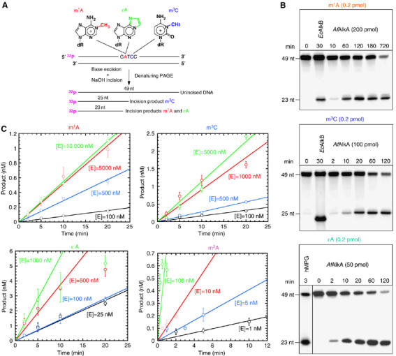
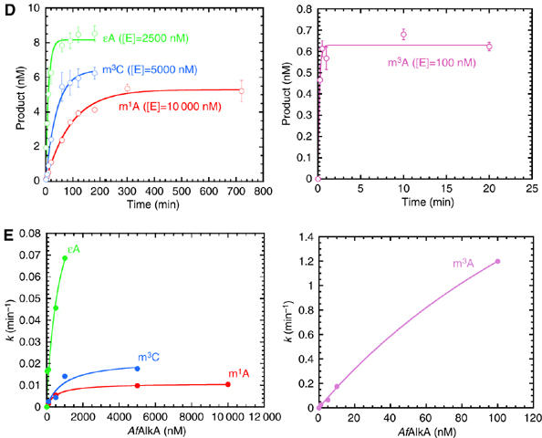
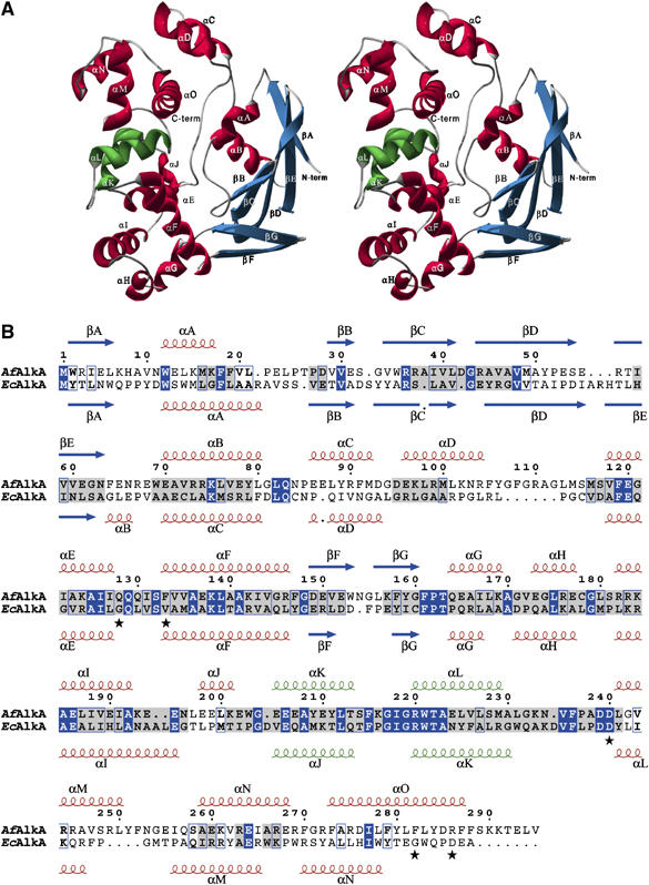
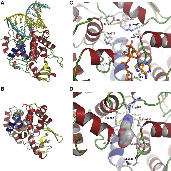
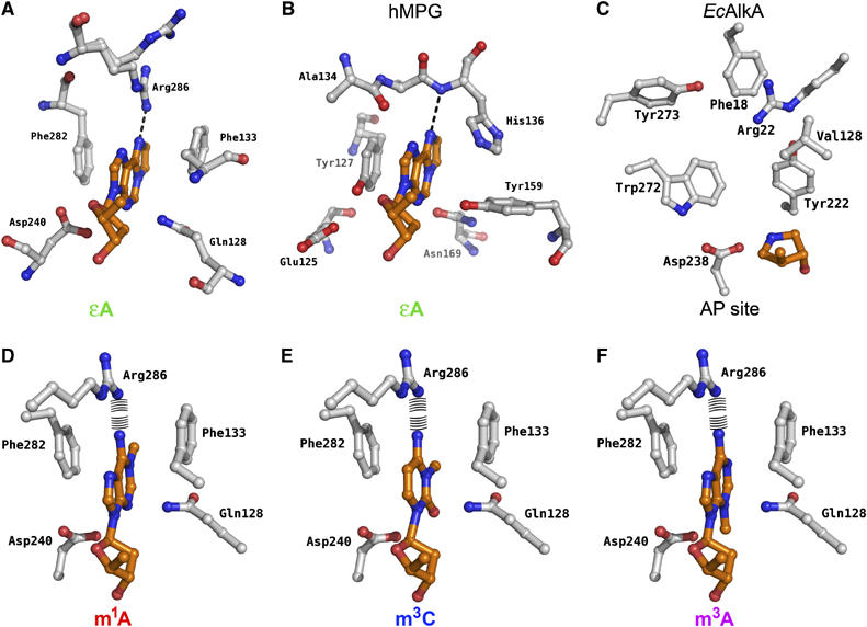
N(1)-methyladenine (m(1)A) and N(3)-methylcytosine (m(3)C) are major toxic and mutagenic lesions induced by alkylation in single-stranded DNA. In bacteria and mammals, m(1)A and m(3)C were recently shown to be repaired by AlkB-mediated oxidative demethylation, a direct DNA damage reversal mechanism. No AlkB gene homologues have been identified in Archaea. We report that m(1)A and m(3)C are repaired by the AfAlkA base excision repair glycosylase of Archaeoglobus fulgidus, suggesting a different repair mechanism for these lesions in the third domain of life. In addition, AfAlkA was found to effect a robust excision of 1,N(6)-ethenoadenine. We present a high-resolution crystal structure of AfAlkA, which, together with the characterization of several site-directed mutants, forms a molecular rationalization for the newly discovered base excision activity.


Figures

Figure 1abc
Figure 1abc
Excision of m1A, m3C and ɛA from DNA by AfAlkA. (A) The oligonucleotides containing m1A, ɛA or m3C at a specific position utilized as substrates. The size of the incision products following glycosylase excision of the specified base lesion and base-catalyzed phosphodiester bond cleavage of the resulting abasic site by alkali treatment is indicated. (B) Cleavage of 5′32P-labeled 49-nucleotides (nt) DNA into repair product (23 and 25 nt) is shown, for typical experiments. Incubation with EcAlkB (4.2 pmol) at 37°C was included as a positive control (in the case of m1A, 10 fmol DNA; m3C, 4.2 fmol DNA), where the DpnII cleavage site corresponds to 22 nt (Ringvoll et al, 2006) (not indicated). For excision of ɛA, incubation of 10 fmol DNA with a 26 kDa truncated hMPG protein (O'Connor, 1993) was included as a positive control. (C) Single-turnover kinetics for excision of m1A, m3C and ɛA, where 10 nM of DNA was incubated with the indicated concentrations of AfAlkA at 70°C for increasing time periods. Incubation with MNU-treated DNA (see Materials and methods) was performed under conditions where virtually only m3A is enzymatically excised (Birkeland et al, 2002), using 5000 d.p.m. of methylated DNA bases (∼1 nM of m3A). Each value represents the average of three independent measurements.
Figure 1de
Figure 1de
(D) Plots of product formation as a function of time ([P]–time plots, together with a curve fit of equation (2) to the experimental data, to determine the active substrate concentration [DNA]tot (see Results)). (E) Calculated k values (determined from the slopes of the curves presented in C) as a function of [E]tot, together with a curve fit of equation (3) to the experimental data (see Results).
Figure 2
Figure 2
Structure of AfAlkA. β-Strands are shown in blue and α-helices in red (except for the two α-helices forming the HhH motif, αK–αL, which are shown in green). (A) Stereo representation of the overall ribbon structure. (B) Primary and secondary structures of AfAlkA (above) aligned with EcAlkA (below). Identical amino-acid residues are boxed in blue, whereas structurally conserved residues are highlighted in gray. AfAlkA residues investigated by site-directed mutagenesis are indicated by an asterisk. The sequence alignment in (B) is based on a structural alignment of the crystal structures. The secondary structure elements of AfAlkA are indicated above the alignment, whereas those of EcAlkA are indicated below.
Figure 3
Figure 3
Crystal structures of (A) EcAlkA in complex with DNA containing the modified abasic nucleotide 1-azaribose (Hollis et al, 2000) and (B) AfAlkA, including an enlarged view of their respective active site regions (C, D). The damage-containing strand is shown in cyan and the complementary strand is shown in yellow. The HhH motif of EcAlkA and AfAlkA is colored dark blue, whereas the amino-acid residue replacing the flipped-out nucleotide is colored red in (A and B). In (D), an ɛA moiety has been modeled into the substrate-binding pocket of AfAlkA, with the molecular surface of the ɛA moiety shown in atom colors. Furthermore, the AfAlkA residue Arg286 is flexible and, as a consequence, was refined in two conformations, indicated in (D).
Figure 4
Figure 4
Estimated electrostatic surface potential of (A) EcAlkA and (B) AfAlkA colored according to electrostatic potential at a contour level of ±7 kBT (blue, positively charged; red, negatively charged). The damage-containing strand is shown in light blue and the complementary strand in yellow. The DNA fragment from the EcAlkA–DNA complex structure has been modeled to fit to the AfAlkA DNA-binding region in (B).
Figure 5
Figure 5
Substrate-binding pockets of (A) AfAlkA with ɛA modeled into the substrate-binding pocket and (B) hMPG crystallized in complex with an ɛA-containing DNA fragment. The ɛA moiety is indicated with orange carbon atoms. The dotted line indicates the potential hydrogen bond between the damaged base and the protein. (C) Binding pocket of EcAlka crystallized in complex with DNA containing 1-azaribose. (DF) AfAlkA binding pockets with m1A, m3C and m3A modeled, otherwise as above. The potential repulsion between Arg286 and the amine of the damaged base is indicated.
Figure 6
Figure 6
Single-turnover kinetics for excision of m1A (A) and ɛA (B) by mutant AfAlkA proteins using the conditions described in Figure 1(A)–(C) and (D). Each value represents the average of 2–4 independent measurements.

References

    1. Aas PA, Otterlei M, Falnes PØ, Vågbø CB, Skorpen F, Akbari M, Sundheim O, Bjørås M, Slupphaug G, Seeberg E, Krokan HE (2003) Human and bacterial oxidative demethylases repair alkylation damage in both RNA and DNA. Nature 421: 859–863 - PubMed
    1. Abner CW, Lau AY, Ellenberger T, Bloom LB (2001) Base excision and DNA binding activities of human alkyladenine DNA glycosylase are sensitive to the base paired with a lesion. J Biol Chem 276: 13379–13387 - PubMed
    1. Alseth I, Osman F, Korvald H, Tsaneva I, Whitby MC, Seeberg E, Bjørås M (2005) Biochemical characterization and DNA repair pathway interactions of Mag1-mediated base excision repair in Schizosaccharomyces pombe. Nucleic Acids Res 33: 1123–1131 - PMC - PubMed
    1. Barrows LR, Magee PN (1982) Nonenzymatic methylation of DNA by S-adenosylmethionine in vitro. Carcinogenesis 3: 349–351 - PubMed
    1. Beranek DT (1990) Distribution of methyl and ethyl adducts following alkylation with monofunctional alkylating agents. Mutat Res 231: 11–30 - PubMed

Publication types