Comparative Study
. 2007 Apr 18;26(8):1995-2004.
doi: 10.1038/sj.emboj.7601661. Epub 2007 Mar 29.

Nanodiscs unravel the interaction between the SecYEG channel and its cytosolic partner SecA

Affiliations
Comparative Study

Nanodiscs unravel the interaction between the SecYEG channel and its cytosolic partner SecA

Meriem Alami et al. EMBO J. .

Abstract

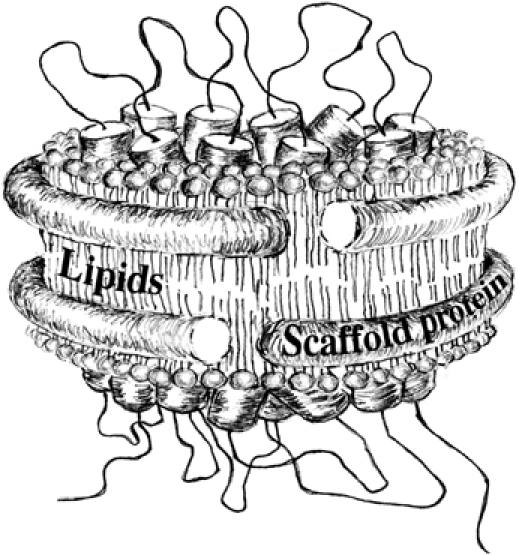
The translocon is a membrane-embedded protein assembly that catalyzes protein movement across membranes. The core translocon, the SecYEG complex, forms oligomers, but the protein-conducting channel is at the center of the monomer. Defining the properties of the SecYEG protomer is thus crucial to understand the underlying function of oligomerization. We report here the reconstitution of a single SecYEG complex into nano-scale lipid bilayers, termed Nanodiscs. These water-soluble particles allow one to probe the interactions of the SecYEG complex with its cytosolic partner, the SecA dimer, in a membrane-like environment. The results show that the SecYEG complex triggers dissociation of the SecA dimer, associates only with the SecA monomer and suffices to (pre)-activate the SecA ATPase. Acidic lipids surrounding the SecYEG complex also contribute to the binding affinity and activation of SecA, whereas mutations in the largest cytosolic loop of the SecY subunit, known to abolish the translocation reaction, disrupt both the binding and activation of SecA. Altogether, the results define the fundamental contribution of the SecYEG protomer in the translocation subreactions and illustrate the power of nanoscale lipid bilayers in analyzing the dynamics occurring at the membrane.


Figures

Figure 1
Figure 1
Schematic view of a membrane protein complex embedded into the Nanodisc structure (adapted from Bayburt and Sligar, 2003). The image was kindly provided by Ms Kailun Jiang.
Figure 2
Figure 2
Reconstitution of the SecYEG complex into Nanodiscs. (A) Typical protein elution profile obtained after size-exclusion chromatography of a SecYEG–Nanodisc preparation. (B) The fractions corresponding to elution volume 10–16 ml in (A) were analyzed by SDS–PAGE, followed by Coomassie blue staining of the gel. For molecular size comparison, the SecYEG complex and the MSPs were loaded on thetwo lanes on the left. (C) Sedimentation velocity analysis and c(s) distribution of the Nd–SecYEG complex at 5.6 μM (gray lane) and 9.6 μM (black lane). A small peak is present at lower and higher S values, which represent small amount of free MSP and MSP bound to a dimeric SecYEG complex, respectively. The insets represent the c(M) distribution of the 9.6-μM sample (f/f0=1.4).
Figure 3
Figure 3
The SecYEG monomer is stably incorporated into the Nanodisc structure. (A) The Nd–SecYEG complex (4 μg, lane 2) was analyzed by BN-PAGE followed by Coomassie blue staining. For comparison, the detergent-soluble SecYEG complex (4 μg, lane 1), the empty discs (2 μg, lane 3) and the MSPs (2 μg, lane 4) were loaded on the same gel. Molecular weight markers: ferritin, 440 kDa; catalase, 232 kDa; aldolase, 158 kDa and BSA 66/132 kDa. (B) The protein samples described in (A) were analyzed by CN-PAGE, followed by Coomassie blue staining of the gel. (C) The [125I]SecYEG and [125I]Nd–SecYEG complexes (each ∼20 000 c.p.m.; ∼20 ng) were incubated at the indicated temperature for 5 min before analysis by BN-PAGE and phosphorimaging. In the presence of 0.1% SDS, both complexes are dissociated.
Figure 4
Figure 4
Binding of the SecA protein onto the SecYEG–Nanodisc. (A) The SecA protein (1 μg, lanes 1–5) was incubated for 5 min at room temperature with the indicated amount of SecYEG–Nanodiscs. As control, the empty discs (2 μg, lanes 6 and 7) were incubated with or without SecA (0.5 μg). The protein samples were separated by CN-PAGE followed by Coomassie blue staining of the gel. (B) The indicated amounts of SecA proteins were incubated with (even lanes) or without (odd lanes) the Nd–SecYEG complex (2 μg) and analyzed by CN-PAGE followed by Coomassie blue staining.
Figure 5
Figure 5
Binding of disulfide-linked SecA onto the SecYEG–Nanodisc. (A) The SecA protein was oxidized with Cu2+(phenanthroline)3 at the indicated concentration and aliquots (1 μg) were analyzed by non-reducing SDS–PAGE, followed by Coomassie blue staining of the gel. (B) The same SecA aliquots (labeled SecACP3 on the figure) were analyzed by CN-PAGE, either alone (lanes 1–4) or after incubation with the Nd–SecYEG complex (2 μg; lanes 5–8). (C) Separation of the Nd–SecYEG–SecA complexes by sucrose density centrifugation. The Nd–SecYEG complex was reconstituted in the presence of trace amounts of 125I-labeled SecYEG. The samples contained (1) 70 μg BSA, 70 μg SecA and 30 μg ferritin, (2) [125I]Nd–SecYEG, (3) [125I]Nd–SecYEG+70 μg SecA and (4) [125I]Nd–SecYEG+70 μg of purified cysteine-linked SecA dimer. Samples from fractions 6–20 were analyzed by SDS–PAGE, followed by Coomassie blue staining (panel 1) or phosphorimaging (panels 2–4). The band detected by phosphorimaging is [125I]SecY. Crosslinked SecA dimer and unmodified SecA dimer sediment at the same position on these sucrose gradients (data not shown).
Figure 6
Figure 6
Crosslinking analysis of the Nd–SecYEG–SecA complex. (A) About 2 μg of [125I]Nd–SecYEG (lane 1) was mixed with 1 μg SecA (lane 2) and then incubated with the crosslinker reagent EGS as described in Materials and methods. In the reverse experiment, [125I]SecA (0.5 μg, lanes 3–6) was incubated with unlabeled Nd–SecYEG (2 μg, lane 5) or empty Nanodiscs (2 μg, lane 6) before crosslinking with EGS. Proteins were dissolved in 0.1% SDS and analyzed by BN-PAGE followed by Coomassie blue staining (left panel) and phosphorimaging (right panel). (B) [125I]SecA (0.5 μg) was incubated with the indicated amounts of Nd–SecYEG or empty Nanodiscs. The crosslinking reaction was performed with EGS as described in Materials and methods.
Figure 7
Figure 7
Analysis of the SecA dimer dissociation by steady-state FRET. Excitation was set at 390 nm and emitted light was recorded from 420 to 580 nm. (A) Coumarin/fluorescein-labeled SecA heterodimer (9 μg) (dark trace) compared with coumarin/unlabeled (dashed dark trace) or fluorescein/unlabeled SecA heterodimer (9 μg) (dashed gray trace). The emitted light for coumarin/fluorescein-labeled SecA heterodimer (9 μg, dark trace) was again recorded after the following addition (gray traces). (B) 45 μg of unlabeled SecA; (C) 45 μg of Nd–SecYEG; and (D) 18 μg of empty Nanodiscs.
Figure 8
Figure 8
The SecY residue R357 and acidic lipids contribute to the binding of SecA. (A) Wild-type or mutant SecYEG Nanodiscs (1 μg) were incubated for 5 min at room temperature with the indicated amounts of SecA. Samples were analyzed by CN-PAGE followed by Coomassie blue staining of the gel. Incubation at 37°C did not change the results (data not shown). (B, C) The SecYEG complex reconstituted into Nanodisc (1 μg) with the indicated phospholipids was incubated for 5 min at room temperature with the indicated amount of SecA protein and then analyzed by CN-PAGE and Coomassie blue staining. Given the smeary aspect of the Nd–SecYEG–SecA complex on native gel, densitometry analysis was performed on unbound SecA obtained at saturating binding concentration (i.e. 2 μg of SecA in this experiment). The densitometry values were compared with SecA concentration standards run on the same gel.
Figure 9
Figure 9
Activation of the SecA ATPase at Nanodisc-reconstituted SecYEG. (A) SecA (1 μg) was incubated at 37°C in TKA buffer (50 mM KCl, 1 mM DTT, 1 mM ATP and 25 mM Tris–HCl pH 8.8) alone or in the presence of 4 μg of either the Nd–SecYEG complex, the empty discs or the MSPs. The amount of inorganic phosphate released during incubation was measured every 5 min by photocolorimetric method. (B) SecA (1 μg) was incubated at 37°C in TKA buffer in the presence of 4 μg of the indicated Nanodisc preparation. The amount of inorganic phosphate was measured after 20 min of incubation. (C) 125I-labeled SecA protein (∼20 000 c.p.m., ∼20 ng, lane 1) was incubated for 5 min at 37°C in TSG buffer with 1 μg of SecYEG–Nanodiscs (lanes 2–11) in the presence or absence of 1 mM ATP. Increasing amounts of unlabeled SecA were then added as indicated for an additional 5 min at 37°C. Protein samples were separated by CN-PAGE and analyzed by phosphorimaging.

References

    1. Bassilana M, Arkowitz RA, Wickner W (1992) The role of the mature domain of proOmpA in the translocation ATPase reaction. J Biol Chem 267: 25246–25250 - PubMed
    1. Bayburt TH, Grinkova YV, Sligar SG (2006) Assembly of single bacteriorhodopsin trimers in bilayer nanodiscs. Arch Biochem Biophys 450: 215–222 - PubMed
    1. Bayburt TH, Sligar SG (2003) Self-assembly of single integral membrane proteins into soluble nanoscale phospholipid bilayers. Protein Sci 12: 2476–2481 - PMC - PubMed
    1. Beckmann R, Spahn CM, Eswar N, Helmers J, Penczek PA, Sali A, Frank J, Blobel G (2001) Architecture of the protein-conducting channel associated with the translating 80S ribosome. Cell 107: 361–372 - PubMed
    1. Bessonneau P, Besson V, Collinson I, Duong F (2002) The SecYEG preprotein translocation channel is a conformationally dynamic and dimeric structure. EMBO J 21: 995–1003 - PMC - PubMed

Publication types

MeSH terms