. 2007 Mar;3(3):e51.
doi: 10.1371/journal.ppat.0030051.

Bacterial ligands generated in a phagosome are targets of the cytosolic innate immune system

Affiliations

Bacterial ligands generated in a phagosome are targets of the cytosolic innate immune system

Anat A Herskovits et al. PLoS Pathog. 2007 Mar.

Abstract

Macrophages are permissive hosts to intracellular pathogens, but upon activation become microbiocidal effectors of innate and cell-mediated immunity. How the fate of internalized microorganisms is monitored by macrophages, and how that information is integrated to stimulate specific immune responses is not understood. Activation of macrophages with interferon (IFN)-gamma leads to rapid killing and degradation of Listeria monocytogenes in a phagosome, thus preventing escape of bacteria to the cytosol. Here, we show that activated macrophages induce a specific gene expression program to L. monocytogenes degraded in the phago-lysosome. In addition to activation of Toll-like receptor (TLR) signaling pathways, degraded bacteria also activated a TLR-independent transcriptional response that was similar to the response induced by cytosolic L. monocytogenes. More specifically, degraded bacteria induced a TLR-independent IFN-beta response that was previously shown to be specific to cytosolic bacteria and not to intact bacteria localized to the phagosome. This response required the generation of bacterial ligands in the phago-lysosome and was largely dependent on nucleotide-binding oligomerization domain 2 (NOD2), a cytosolic receptor known to respond to bacterial peptidoglycan fragments. The NOD2-dependent response to degraded bacteria required the phagosomal membrane potential and the activity of lysosomal proteases. The NOD2-dependent IFN-beta production resulted from synergism with other cytosolic microbial sensors. This study supports the hypothesis that in activated macrophages, cytosolic innate immune receptors are activated by bacterial ligands generated in the phagosome and transported to the cytosol.


Conflict of interest statement

Competing interests. DAP has a consulting relationship with and a financial interest in Cerus Corporation, which stands to benefit from commercialization of the results of this research.

Figures

Figure 1
Figure 1. L. monocytogenes Is Killed and Degraded in IFN-γ–Activated Macrophages, Leading to a Specific Activation Program
(A) Intracellular growth curve of w.t. L. monocytogenes (L.m.) and LLO-minus mutant (LLO-) in IFN-γ–activated (•) versus non-activated () resident peritoneal macrophages. (B) Intracellular growth curve of w.t. L. monocytogenes and LLO-minus mutant in IFN-γ–activated (•) versus non-activated () BMD macrophages. (C) Immunofluorescence microscopy of BMD macrophages infected with GFP-expressing, w.t. L. monocytogenes and LLO-minus mutant at 4 h.p.i. Macrophage actin was stained in red with rhodamine-phalloidin. Lower panel, staining of acidic vesicles with LysoTracker RED in LLO-minus and w.t. L. monocytogenes infections (w.t. infection is also stained with cuomarin-phalloidin for actin labeling in blue). Immunofluorescence data are representative of 90% of the infected macrophages in two independent experiments. (D) Macrophages transcriptional response to w.t. L. monocytogenes and LLO-minus mutant infection with and without activation of IFN-γ. Only the most changed genes upon bacterial infection of non-activated macrophages are presented (253 genes vary 5.6-fold and up). Genes were clustered using Pearson hierarchical clustering. All columns were normalized to uninfected macrophages (un) without IFN-γ treatment. Rows colorimetrically represent expression ratios of individual genes as describe in material and methods. (E) Presentation of selected genes (cytosolic-induced, phagosomal-induced, and both) and their expression profile in activated and non-activated macrophages infected with w.t. L. monocytogenes and LLO-minus bacteria.
Figure 2
Figure 2. IFN-β Expression by LLO-Minus Mutant Is independent of TLR3, 7, and 9
(A) Time course analysis of ifnβ induction by w.t. L. monocytogenes (L.m.) and LLO-minus mutant (LLO-) in non-activated and IFN-γ–activated BMD macrophages. Analysis of IFN-β mRNA by Q-RT-PCR at 2, 4, and 6 h.p.i. un, uninfected macrophages. (B) Analysis of ifnβ induction in response to the LLO-minus mutant after infection of MyD88- and Trif-deficient activated BMD macrophages by Q-RT-PCR at 6 h.p.i. (C) Immunofluorescence microscopy of MyD88 and C57BL/6 activated BMD macrophages infected with the GFP-expressing LLO-minus mutant at 6 h.p.i. (D) Analysis of ifnβ induction in response to the LLO-minus mutant after infection of TLR3-, 7-, and 9–deficient activated BMD macrophages by Q-RT-PCR. Data correspond to the mean ± standard error of the mean (s.e.m.) (triplicate determinations) and are representative of three or more independent experiments. Immunofluorescence data are representative of 90% of the infected macrophages in two independent experiments.
Figure 3
Figure 3. Role of NOD2 in the IFN-β Response to L. monocytogenes in Activated Macrophages
(A) Analysis of ifnβ induction by Q-RT-PCR and IFN-β secretion (ELISA) by L. monocytogenes (L.m.) and the LLO-minus mutant (LLO-) in activated, NOD2-, and NOD1-deficient BMD macrophages. The level of induction in NOD2−/− was compared to macrophages from w.t. NOD2 mice littermates. un, uninfected macrophages. (B) Analysis of ifnβ induction (Q-RT-PCR) by L. monocytogenes and the LLO-minus mutant in activated, IRF-3–deficient BMD macrophages in comparison to C57BL/6 macrophages. un, uninfected macrophages. (C) ELISA analysis of IFN-β amounts secreted by NOD2-deficient BMD macrophages in response to 100 μg/ml MDP and 100 μg/ml poly(I:C) delivered with lipofectamine. Data correspond to the mean ± s.e.m (triplicate determinations) and are representative of three or more independent experiments.
Figure 4
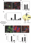
Figure 4. Effects of Phagosome Acidification and Phagosomal Membrane Potential on IFN-β Expression
(A) Immunofluorescence microscopy of C57BL/6 BMD macrophages infected with GFP-expressing LLO-minus mutant (LLO-) with and without bafilomycin A treatment (6 h.p.i.). (B) Q-RT-PCR analysis of ifnβ induction in response to L. monocytogenes (L.m.) and the LLO-minus mutant with and without bafilomycin A treatment (BafA). Bafilomycin A was added 30 min post-infection. (C) Effect of the ionophors monensin, nigericin, and valinomycin on IFN-β response to the LLO-minus mutant, analyzed by Q-RT-PCR. Schematic presentation of the phagosome, ionophor mode of action, and H+ and K+ ion transfer across the phagosomal membrane. (a.) V-ATPase, which generates an electrochemical potential (ψ), inside positive and acidic. (b.) Monensin or nigericin, which establish a chemical K+ gradient by electroneutral H+/K+ exchange (c.) Valinomycin, which dissipates the electrical potential by electrogenic efflux of K+. (D) Immunofluorescence microscopy of C57BL/6 BMD macrophages infected with GFP-expressing LLO-minus mutant under ionophor treatments. All ionophors were added 30 min post-infection. (E) Effect of monensin (Mon) and chymostatin (Chym) on the IFN-β and IL-12p40 response to LLO-minus mutant in NOD2-deficient cells and NOD2 w.t. cells, analyzed by Q-RT-PCR. Data correspond to the mean ± s.e.m. (triplicate determinations) and are representative of three or more independent experiments. Immunofluorescence data are representative for 99% of the infected macrophages in two independent experiments.
Figure 5
Figure 5. L. monocytogenes PGN Is Degraded in the Phago-Lysosome and Induces NOD2-Dependent Responses
(A) Immunofluorescence microscopy of activated and non-activated BMD macrophages infected with FL-Van–labeled LLO-minus mutant. FL-Van labeling localized to bacterial poles (arrowheads and magnified picture). (B) Response of activated macrophages to L. monocytogenes CW preparation and CW degradation fragments (L.m.CW fragments) delivered with lipofectamine. Level of TNFα, IL-6, and IFN-β induction determined by Q-RT-PCR. (C) Induction of IFN-β response by L. monocytogenes CW fragments in Myd88/Trif double-knockout activated macrophages. Data correspond to the mean ± s.e.m. (triplicate determinations).

References

    1. Stuart LM, Ezekowitz RA. Phagocytosis: Elegant complexity. Immunity. 2005;22:539–550. - PubMed
    1. Schroder K, Hertzog PJ, Ravasi T, Hume DA. Interferon-gamma: An overview of signals, mechanisms and functions. J Leukoc Biol. 2004;75:163–189. - PubMed
    1. Medzhitov R. Toll-like receptors and innate immunity. Nat Rev Immunol. 2001;1:135–145. - PubMed
    1. Inohara, Chamaillard, McDonald C, Nunez G. NOD-LRR proteins: Role in host-microbial interactions and inflammatory disease. Annu Rev Biochem. 2005;74:355–383. - PubMed
    1. Rutz M, Metzger J, Gellert T, Luppa P, Lipford GB, et al. Toll-like receptor 9 binds single-stranded CpG-DNA in a sequence- and pH-dependent manner. Eur J Immunol. 2004;34:2541–2550. - PubMed

Publication types

MeSH terms