

Measurement of collagen and smooth muscle cell content in atherosclerotic plaques using polarization-sensitive optical coherence tomography
- PMID: 17397678
- PMCID: PMC2785549
- DOI: 10.1016/j.jacc.2006.11.040
Measurement of collagen and smooth muscle cell content in atherosclerotic plaques using polarization-sensitive optical coherence tomography
Abstract
Objectives: The purpose of this study was to investigate the measurement of collagen and smooth muscle cell (SMC) content in atherosclerotic plaques using polarization-sensitive optical coherence tomography (PSOCT).
Background: A method capable of evaluating plaque collagen content and SMC density can provide a measure of the mechanical fidelity of the fibrous cap and can enable the identification of high-risk lesions. Optical coherence tomography has been demonstrated to provide cross-sectional images of tissue microstructure with a resolution of 10 mum. A recently developed technique, PSOCT measures birefringence, a material property that is elevated in tissues such as collagen and SMCs.
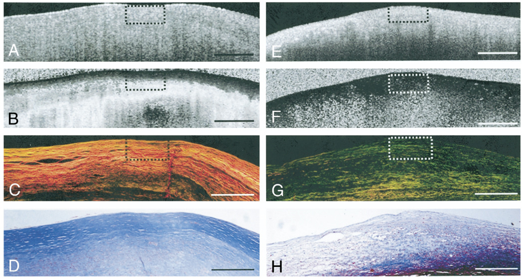
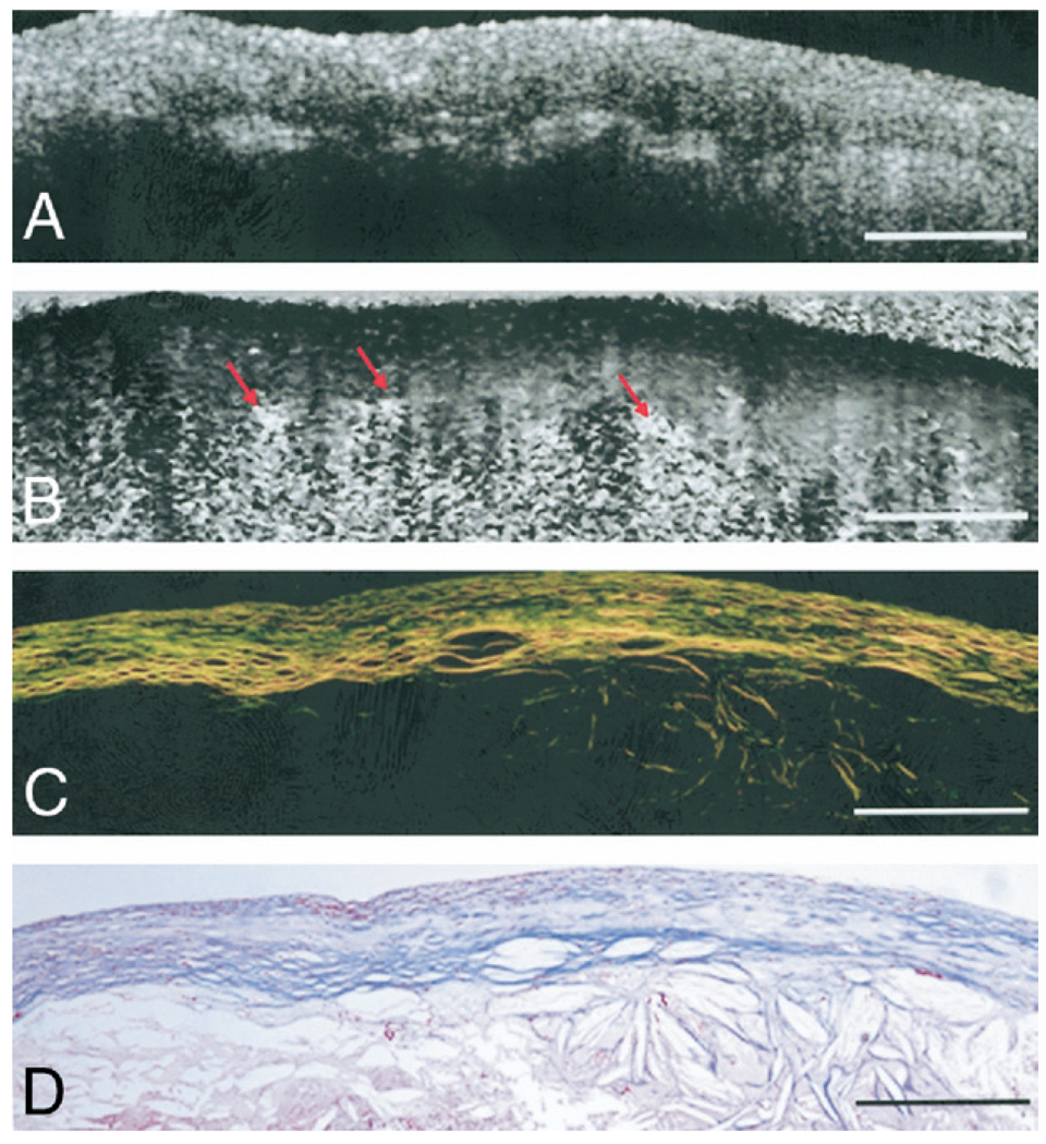
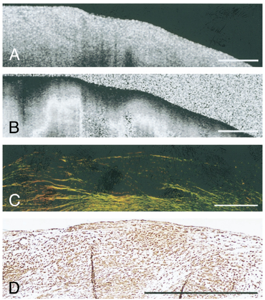
Methods: We acquired PSOCT images of 87 aortic plaques obtained from 20 human cadavers. Spatially averaged PSOCT birefringence, Phi, was measured and compared with plaque collagen and SMC content, quantified morphometrically by picrosirius red and smooth muscle actin staining at the corresponding locations.
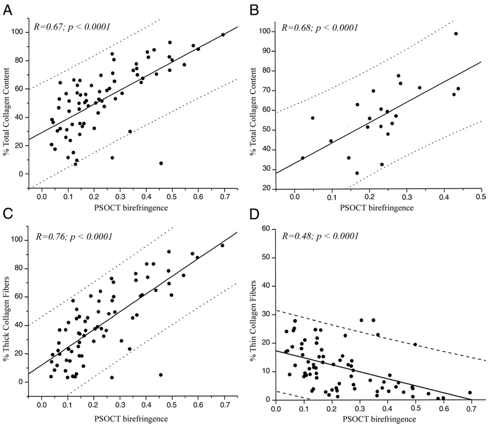
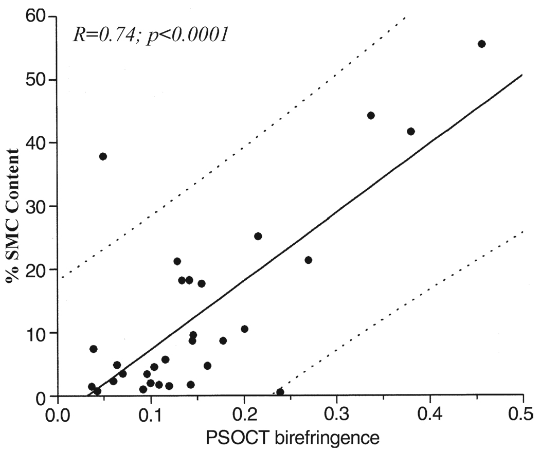
Results: There was a high positive correlation between PSOCT measurements of Phi and total collagen content in all plaques (r = 0.67, p < 0.001) and in fibrous caps of necrotic core fibroatheromas (r = 0.68, p < 0.001). Polarization-sensitive optical coherence tomography measurements of Phi demonstrated a strong positive correlation with thick collagen fiber content (r = 0.76, p < 0.001) and SMC density (r = 0.74, p < 0.01).
Conclusions: Our results demonstrate that PSOCT enables the measurement of birefringence in plaques and in fibrous caps of necrotic core fibroatheromas. Given its potential to evaluate collagen content, collagen fiber thickness, and SMC density, we anticipate that PSOCT will significantly improve our ability to evaluate plaque stability in patients.
Figures

References
-
- Arroyo LH, Lee RT. Mechanisms of plaque rupture: mechanical and biologic interactions. Cardiovasc Res. 1999;41:369–375. - PubMed
-
- Falk E, Shah PK, Fuster V. Coronary plaque disruption. Circulation. 1995;92:657–671. - PubMed
-
- Virmani R, Kolodgie FD, Burke AP, Farb A, Schwartz SM. Lessons from sudden coronary death: a comprehensive morphological classification scheme for atherosclerotic lesions. Arterioscler Thromb Vasc Biol. 2000;20:1262–1275. - PubMed
-
- Bauriedel G, Hutter R, Welsch U, Bach R, Sievert H, Luderitz B. Role of smooth muscle cell death in advanced coronary primary lesions: implications for plaque instability. Cardiovasc Res. 1999;41:480–488. - PubMed
-
- Newby AC, Zaltsman AB. Fibrous cap formation or destruction—the critical importance of vascular smooth muscle cell proliferation, migration and matrix formation. Cardiovasc Res. 1999;41:345–360. - PubMed
Publication types
MeSH terms
Substances
Grants and funding
LinkOut - more resources
Full Text Sources
Other Literature Sources
Medical
Miscellaneous
