Review
. 2007 May 22;581(11):2140-9.
doi: 10.1016/j.febslet.2007.03.032. Epub 2007 Mar 21.

Cellular functions of NSF: not just SNAPs and SNAREs

Affiliations
Review

Cellular functions of NSF: not just SNAPs and SNAREs

Chunxia Zhao et al. FEBS Lett. .

Abstract

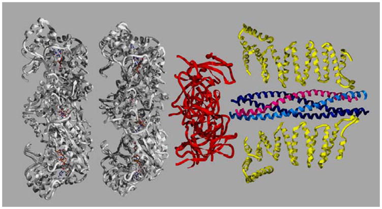
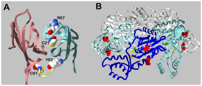
N-ethylmaleimide sensitive factor (NSF) is an ATPases associated with various cellular activities protein (AAA), broadly required for intracellular membrane fusion. NSF functions as a SNAP receptor (SNARE) chaperone which binds, through soluble NSF attachment proteins (SNAPs), to SNARE complexes and utilizes the energy of ATP hydrolysis to disassemble them thus facilitating SNARE recycling. While this is a major function of NSF, it does seem to interact with other proteins, such as the AMPA receptor subunit, GluR2, and beta2-AR and is thought to affect their trafficking patterns. New data suggest that NSF may be regulated by transient post-translational modifications such as phosphorylation and nitrosylation. These new aspects of NSF function as well as its role in SNARE complex dynamics will be discussed.


Figures

Figure 1
Figure 1. 20S Particle
Depicted are the crystal structures for each element of the 20S particle. The two ATP-binding domains of NSF (D1 and D2) in white are modeled from the NSF-D2 structure (1D2N). A trimer of NSF-N domains, in red, is based on the three-in-three-out model of May et al. 1999 (1QDN). Only two yeast α-SNAPs (Sec17p, 1QQE ) are depicted in yellow. The coiled-coil SNARE complex is depicted with syntaxin-1a in light blue, synaptobrevin/VAMP-2, in magenta, and SNAP-25 in dark blue (2BUO). The images were created with Swiss PDB viewer and rendered with Pov-Ray.
Figure 2
Figure 2. Post-translational modifications of NSF
Panel A, crystal structure of the amino-terminal domain of NSF, showing the locations of the modified cysteine and tyrosine residues. The NA subdomain is in rose and the NB subdomain is in aqua. The image is based on 1QDN and was generated using Swiss PDB viewer and rendered with Pov-Ray. Panel B, crystal structure of the NSF-D2 hexamer. Three subunits are shown in white, two in aqua and one in blue. The modified residues are indicated by red. Position 1 is predicted to be equivalent to the phosphorylated Ser237 in the NSF-D1 domain. Position 2 is Ser569, phosphorylated by Pctaire1. The image is based on 1D2N and was created with Swiss PDB viewer and rendered with Pov-Ray.

References

    1. Block MR, Glick BS, Wilcox CA, Wieland FT, Rothman JE. Purification of an N-ethylmaleimide-sensitive protein catalyzing vesicular transport. Proc Natl Acad Sci U S A. 1988;85:7852–6. - PMC - PubMed
    1. Clary DO, Griff IC, Rothman JE. SNAPs, a family of NSF attachment proteins involved in intracellular membrane fusion in animals and yeast. Cell. 1990;61:709–21. - PubMed
    1. Sollner T, Whiteheart SW, Brunner M, Erdjument-Bromage H, Geromanos S, Tempst P, Rothman JE. SNAP receptors implicated in vesicle targeting and fusion. Nature. 1993;362:318–24. - PubMed
    1. Weber T, Zemelman BV, McNew JA, Westermann B, Gmachl M, Parlati F, Sollner TH, Rothman JE. SNAREpins: minimal machinery for membrane fusion. Cell. 1998;92:759–72. - PubMed
    1. Fasshauer D, Sutton RB, Brunger AT, Jahn R. Conserved structural features of the synaptic fusion complex: SNARE proteins reclassified as Q- and R-SNAREs. Proc Natl Acad Sci U S A. 1998;95:15781–6. - PMC - PubMed

Publication types

MeSH terms

Substances

LinkOut - more resources