

Prenatal choline deficiency increases choline transporter expression in the septum and hippocampus during postnatal development and in adulthood in rats
- PMID: 17399691
- PMCID: PMC1952662
- DOI: 10.1016/j.brainres.2007.03.004
Prenatal choline deficiency increases choline transporter expression in the septum and hippocampus during postnatal development and in adulthood in rats
Abstract
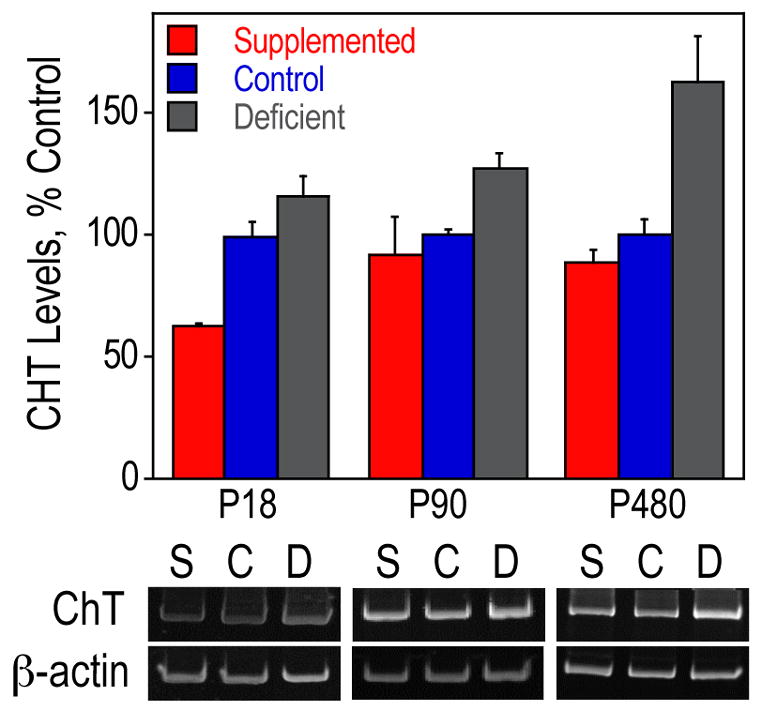
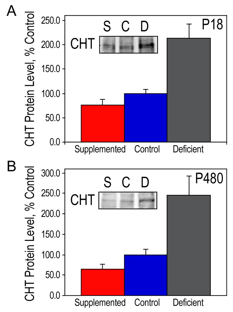
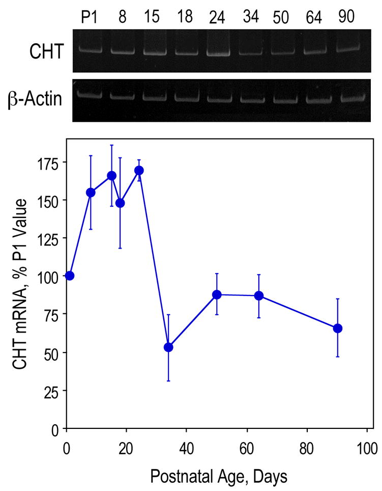
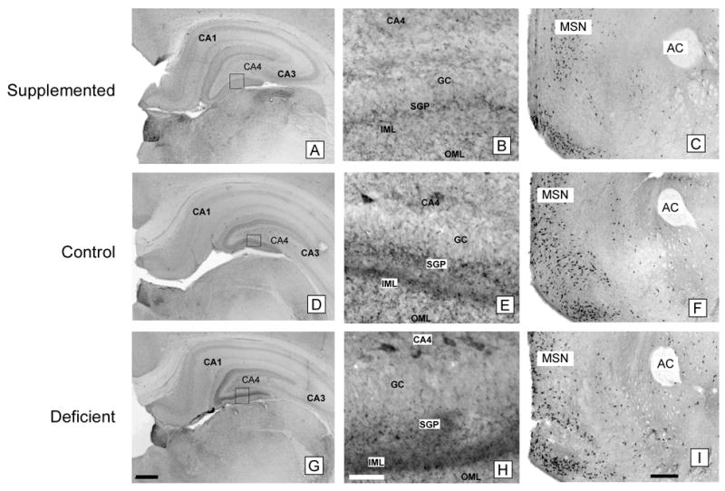
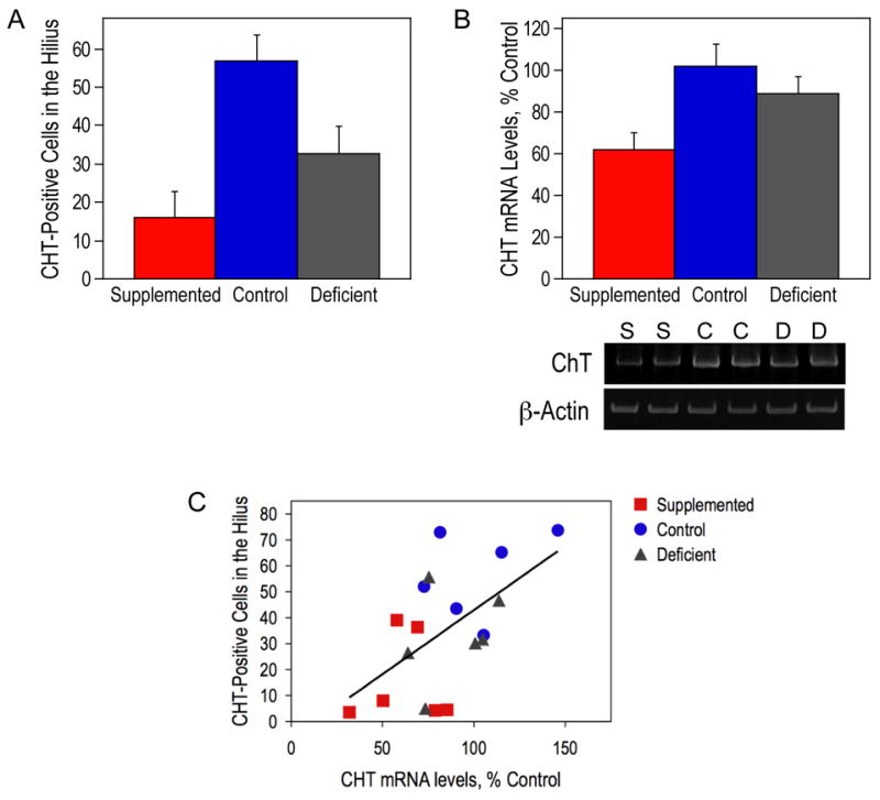
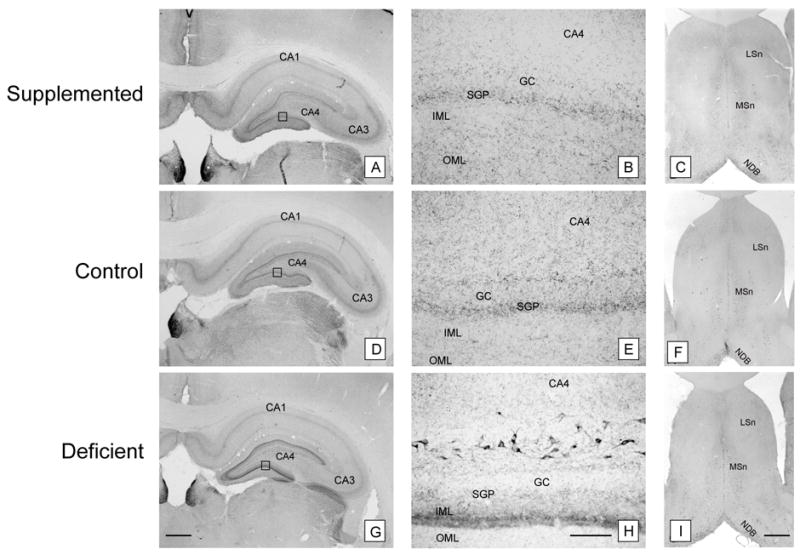
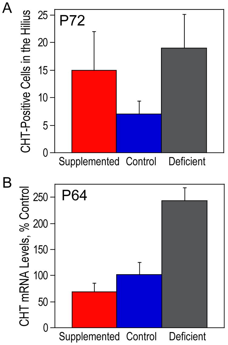
Supplementation of maternal diet with the essential nutrient, choline, during the second half of pregnancy in rats causes long-lasting improvements in spatial memory in the offspring and protects them from the memory decline characteristic of old age. In contrast, prenatal choline deficiency is associated with poor performance in certain cognitive tasks. The mechanism by which choline influences learning and memory remains unclear; however, it may involve changes to the hippocampal cholinergic system. Previously, we showed that the hippocampi of prenatally [embryonic days (E) 11-17] choline-deficient animals have increased synthesis of acetylcholine (ACh) from choline transported by the high-affinity choline transporter (CHT) and reduced ACh content relative to the control and to the E11-17 choline-supplemented rats. In the current study, we found that, during postnatal period [postnatal days (P) 18-480], prenatal choline deficiency increased the expression of CHT mRNA in the septum and CHT mRNA and protein levels in the hippocampus and altered the pattern of CHT immunoreactivity in the dentate gyrus. CHT immunoreactivity was more prominent in the inner molecular layer in prenatally choline-deficient rats compared to controls and prenatally choline-supplemented animals. In addition, in all groups, we observed a population of hilar interneurons that were CHT-immunoreactive. These neurons are the likely source of the hippocampal CHT mRNA as their number correlated with the levels of this mRNA. The abundance of hippocampal CHT mRNA rose between P1 and P24 and then declined reaching 60% of the P1 value by P90. These data show that prenatal availability of choline alters its own metabolism (i.e., CHT expression). While the upregulated CHT expression during the period of prenatal choline deficiency may be considered as a compensatory mechanism that could enhance ACh synthesis when choline supply is low, the persistent upregulation of CHT expression subsequent to the brief period of prenatal deprivation of choline in utero might be beneficial during choline deficiency in adulthood.
Figures
Similar articles
-
Prenatal choline supplementation in rats increases the expression of IGF2 and its receptor IGF2R and enhances IGF2-induced acetylcholine release in hippocampus and frontal cortex.Brain Res. 2008 Oct 27;1237:124-35. doi: 10.1016/j.brainres.2008.08.046. Epub 2008 Aug 28. Brain Res. 2008. PMID: 18786520
-
Prenatal availability of choline alters the development of acetylcholinesterase in the rat hippocampus.Dev Neurosci. 1999;21(2):94-104. doi: 10.1159/000017371. Dev Neurosci. 1999. PMID: 10449981
-
Prenatal choline availability modulates hippocampal and cerebral cortical gene expression.FASEB J. 2007 May;21(7):1311-23. doi: 10.1096/fj.06-6597com. Epub 2007 Jan 30. FASEB J. 2007. PMID: 17264169
-
Prenatal choline deficiency does not enhance hippocampal vulnerability after kainic acid-induced seizures in adulthood.Brain Res. 2011 Sep 21;1413:84-97. doi: 10.1016/j.brainres.2011.07.042. Epub 2011 Jul 24. Brain Res. 2011. PMID: 21840511 Free PMC article.
-
The supply of choline is important for fetal progenitor cells.Semin Cell Dev Biol. 2011 Aug;22(6):624-8. doi: 10.1016/j.semcdb.2011.06.002. Epub 2011 Jun 12. Semin Cell Dev Biol. 2011. PMID: 21693194 Free PMC article. Review.
Cited by
-
Prenatal choline availability alters the context sensitivity of Pavlovian conditioning in adult rats.Learn Mem. 2008 Dec 2;15(12):866-75. doi: 10.1101/lm.1058708. Print 2008 Dec. Learn Mem. 2008. PMID: 19050158 Free PMC article.
-
Prenatal choline supplementation increases sensitivity to contextual processing of temporal information.Brain Res. 2008 Oct 27;1237:204-13. doi: 10.1016/j.brainres.2008.08.072. Epub 2008 Sep 4. Brain Res. 2008. PMID: 18778696 Free PMC article.
-
Modulation of Dietary Choline Uptake in a Mouse Model of Acid Sphingomyelinase Deficiency.Int J Mol Sci. 2023 Jun 5;24(11):9756. doi: 10.3390/ijms24119756. Int J Mol Sci. 2023. PMID: 37298714 Free PMC article.
-
The Impact of Traditional Food and Lifestyle Behavior on Epigenetic Burden of Chronic Disease.Glob Chall. 2017 Oct 27;1(8):1700043. doi: 10.1002/gch2.201700043. eCollection 2017 Nov 16. Glob Chall. 2017. PMID: 31565292 Free PMC article. Review.
-
Effects of Maternal Choline Supplementation on the Septohippocampal Cholinergic System in the Ts65Dn Mouse Model of Down Syndrome.Curr Alzheimer Res. 2016;13(1):84-96. doi: 10.2174/1567205012666150921100515. Curr Alzheimer Res. 2016. PMID: 26391045 Free PMC article.
References
-
- Bazalakova MH, Wright J, Schneble EJ, McDonald MP, Heilman CJ, Levey AI, Blakely RD. Deficits in acetylcholine homeostasis, receptors and behaviors in choline transporter heterozygous mice. Genes Brain Behav 2006 - PubMed
-
- Beeri R, Le Novère N, Mervis R, Huberman T, Grauer E, Changeux JP, Soreq H. Enhanced hemicholinium binding and attenuated dendrite branching in cognitively impaired acetylcholinesterase-transgenic mice. J Neurochem. 1997;69:2441–2451. - PubMed
-
- Berse B, Szczecinska W, Lopez-Coviella I, Madziar B, Zemelko V, Kaminski R, Kozar K, Lips KS, Pfeil U, Blusztajn JK. Expression of high affinity choline transporter during mouse development in vivo and its upregulation by NGF and BMP-4 in vitro. Brain Res Dev Brain Res. 2005;157:132–40. - PubMed
-
- Bieri JG. Second report of the ad hoc committee on standards for nutritional studies. J Nutr. 1980;110:1726. - PubMed
-
- Bieri JG, Stoewsand GS, Briggs GM, Phillips RW, Woodard JC, Kanapka JJ. Report of the American Institute of Nutrition ad hoc committee on standards for nutritional studies. J Nutr. 1977;107:1340–1348. - PubMed
Publication types
MeSH terms
Substances
Grants and funding
LinkOut - more resources
Full Text Sources
