. 2007 May 25;369(1):41-54.
doi: 10.1016/j.jmb.2007.03.006. Epub 2007 Mar 12.

Stable complexes formed by HIV-1 reverse transcriptase at distinct positions on the primer-template controlled by binding deoxynucleoside triphosphates or foscarnet

Affiliations

Stable complexes formed by HIV-1 reverse transcriptase at distinct positions on the primer-template controlled by binding deoxynucleoside triphosphates or foscarnet

Peter R Meyer et al. J Mol Biol. .

Abstract

Binding of the next complementary dNTP by the binary complex containing HIV-1 reverse transcriptase (RT) and primer-template induces conformational changes that have been implicated in catalytic function of RT. We have used DNase I footprinting, gel electrophoretic mobility shift, and exonuclease protection assays to characterize the interactions between HIV-1 RT and chain-terminated primer-template in the absence and presence of various ligands. Distinguishable stable complexes were formed in the presence of foscarnet (an analog of pyrophosphate), the dNTP complementary to the first (+1) templating nucleotide or the dNTP complementary to the second (+2) templating nucleotide. The position of HIV-1 RT on the primer-template in each of these complexes is different. RT is located upstream in the foscarnet complex, relative to the +1 complex, and downstream in the +2 complex. These results suggest that HIV-1 RT can translocate along the primer-template in the absence of phosphodiester bond formation. The ability to form a specific foscarnet complex might explain the inhibitory properties of this compound. The ability to recognize the second templating nucleotide has implications for nucleotide misincorporation.


Figures

Figure 1
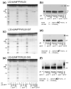
Figure 1. Effects of the next complementary dNTP on DNase I protection and stable complex formation by HIV-1 RT
(a) DNase I footprinting on the 3′-labeled primer strand. Five nM 3'-[32P]ddAMP-terminated L32 primer annealed to WL50 template was incubated in the absence or presence of 200 nM HIV-1 RT and the indicated concentrations of dTTP followed by 3 min exposure to DNase I. Products were separated by electrophoresis through a 20% denaturing polyacrylamide gel. Numbers to the left of the lanes indicate the positions of the bands. The 3'-terminal nucleotide on the primer is defined as position −1 in this and subsequent figures. The arrow indicates the position of the hypersensitive site induced by dTTP binding formed by cleavage between position −18 and −19 on the primer. A partial sequence of the P/T is shown. The +1 base on the template is bold and underlined. AH indicates ddA. (b) EMSA detection of stable complex formation. Labeled P/T was incubated with HIV-1 RT as described in (a) in the absence of dTTP or with the indicated concentrations of dTTP. Incubation at 37°C was followed by addition of heparin/loading dye and separation of DEC from free P/T by electrophoresis through an 8% non-denaturing polyacrylamide gel. (c) Quantitative comparison of primer DNase 1 footprinting and stable complex data as a function of dTTP concentration. The radioactivity in bands at −20 (protected) and −18 (hypersensitive) in (a) and DEC formation in (b) were quantified by phosphorimaging, plotted versus dTTP concentration, and fitted to single-ligand binding curves (solid lines). (d) DNase I footprinting on the 3′-labeled template strand. DNase I footprinting was performed as in (a) but with 3'-[32P]ddAMP-labeled WL50 template annealed with unlabeled ddAMP-terminated L32 primer. The arrow indicates the position of the hypersensitive site induced by dTTP binding formed by cleavage between position −21 and −22 on the template. A partial sequence of the P/T is shown. The +1 base on the template is bold and underlined. AH indicates ddA. (e) Quantitative comparison of template DNase 1 footprinting and stable complex data. The radioactivity in the bands at −7 (protected) and −22 (hypersensitive) in (d) were quantified by phosphorimaging, plotted versus dTTP concentration, and fitted to single-ligand binding curves (solid lines). DEC formation with labeled template is also shown. (f) Kd,app's calculated from data from experiments such as those in (c) and (e). The mean and range are shown for at least two experiments for each value.
Figure 2
Figure 2. Effects of noncomplementary dNTPs on DNase I footprints and stable complex formation by HIV-1 RT
(a), (c), (e) Five nM 3'-[32P] ddAMP-terminated L32 primer annealed to the indicated template was incubated in the absence or presence of HIV-1 RT and the indicated concentrations of dATP, dCTP, dGTP or dTTP followed by exposure to DNase I and electrophoresis performed as in Figure 1(a). Band positions are indicated as in Figure 1. The black and white arrows indicate the hypersensitive sites observed for +1 and +2 complexes, respectively. (b), (d), (f) EMSA detection of stable complex formation. Labeled P/Ts were incubated with HIV-1 RT as in (a),(c), and (e), and the concentrations of dNTPs indicated at the bottom of each panel followed by separation of P/T and DEC as described in Figure 1(b). A partial sequence of each P/T is shown in panels (b), (d) and (f) with the +1 and +2 template nucleotides shown in bold. The +1 nucleotide is underlined. AH indicates ddA .
Figure 3
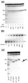
Figure 3. Effects of foscarnet on DNase I protection and stable complex formation by HIV-1 RT
(a) DNase I footprinting as a function of added foscarnet. Labeled 3′-[32P]ddAMP-L32/WL50 P/T was incubated in the absence or presence of HIV-1 RT and the indicated concentrations of foscarnet (fosc) followed by exposure to DNase I and electrophoresis performed as in Figure 1(a). Band positions relative to the primer terminus are shown on the left. The arrows indicate the hypersensitive sites induced by foscarnet. (b) EMSA detection of stable complex formation as a function of foscarnet concentration. Labeled P/T was incubated with HIV-1 RT and the indicated concentrations of foscarnet followed by separation of P/T and DEC as described in the legend to Figure 1(b). (c) Comparison of DNase I footprints with 3′-labeled primer with 800 μM dGTP, 800 μM dTTP or 3.2 mM foscarnet. Labeled P/T was incubated in the absence or presence of HIV-1 RT with the indicated ligand followed by exposure to DNase I and electrophoresis performed as in Figure 1(a). Arrows indicate hypersensitive sites induced by foscarnet (dashed), dTTP (solid black), or dGTP (white).
Figure 4
Figure 4. DNase I protection on P/Ts terminated with dT analogues
Five nM L32 primer annealed to WL50 template was extended with [32P]-dATP, terminated with the indicated dT-analogue and incubated in the absence or presence of 200 nM HIV-1 RT and no ligand, 3.2 mM foscarnet or 200 μM of the indicated dNTP followed by exposure to DNase I and electrophoresis performed as described in Figure 1(a). Band position relative to the primer terminus is indicated to the left of the lanes. The arrows indicate hypersensitive sites induced by foscarnet (dashed) or +1 dNTP (solid). A partial sequence of the P/T is shown where the first downstream templating base is in bold and underlined. TH indicates the chain-terminating dT-analogue residue in each of the primers (identified at the top of each panel).
Figure 5
Figure 5. Lambda exonuclease mapping of the upstream borders of HIV-1 RT bound to P/T
(a) Five nM 5'-phosphorylated, 3'-[32P]ddAMP-labeled L32 primer annealed to WL50 template was incubated in the absence or presence of excess HIV-1 RT and the indicated ligand followed by incubation with lambda exonuclease. Products were separated by electrophoresis through a non-denaturing 20% polyacrylamide gel and the radioactivity visualized through phosphorimaging. The numbers to the left of the lanes indicate the size of the digestion products (in nucleotides). These values also correspond to positions relative to the primer terminus (e.g., 10 indicates the product of digestion that stopped at the bond upstream of position −10, etc.) Arrows on the right of (a) and at the bottom of panel (c) indicate the predominant stop site for lambda exonuclease digestion in the foscarnet complex (red), +1 complex (green) or +2 complex (blue). (b) The radioactivity was quantified using phosphorimaging for digestion products obtained in the presence of foscarnet (short dashes), dTTP (solid line) or dGTP (long dashes) and plotted versus primer position. (c) Diagram showing map positions for predominant barriers to digestion initiated at the phosphorylated 5′ end of the 3′-labeled primer by lambda exonuclease (shown by the Pac-Man figure). Overlapping colored rectangles represent expected exonuclease protected regions for each complex. The light blue line corresponds to a weaker barrier at position −28 encountered by lambda exonuclease digestion in the presence of dGTP. Nucleotide positions relative to the primer terminus are shown in the scale at the top of the diagram.
Figure 6
Figure 6. RecJf exonuclease mapping of the downstream borders of HIV-1 RT bound to chain-terminated P/T
(a) Five nM 3'-[32P]ddAMP-labeled WL65 template annealed to unlabeled ddAMP-terminated L32 primer was incubated in the absence or presence of 12.5 nM HIV-1 RT and the indicated ligand (0.8 mM dNTP, 3.2 mM foscarnet or no ligand), followed by incubation with RecJf exonuclease as described under Experimental Procedures and electrophoresis performed as in Figure 5. Numbers on the left of the lanes indicate the size of the digestion products (in nucleotides) and the numbers to the right of the lanes indicate the map positions of the predominant downstream barriers to digestion (+1 is the first template base following the primer terminus). The arrows in (a) and (c) indicate predominant barriers to RecJf exonuclease digestion (color-coded as in Figure 5). (b) The radioactivity in the gel in (a) was quantified using phosphorimaging for the products of digestion of complexes formed with foscarnet (short dashes), dTTP (solid line) or dGTP (long dashes) and plotted versus distance from the primer terminus. (c) Diagram showing map positions for predominant barriers to digestion initiated at the 5′ end of the 3′-labeled template by RecJf exonuclease (shown by the Pac-Man figure). Overlapping rectangles represent regions of the P/T protected from exonuclease digestion for each complex. The dark and light red lines correspond to barriers (positions +7 and +8, respectively) encountered by RecJf digestion in the presence of foscarnet. Nucleotide positions relative to the primer terminus are shown in the scale at the bottom of the diagram.

Similar articles

Cited by

References

    1. Johnson KA. Conformational coupling in DNA polymerase fidelity. Annu. Rev. Biochem. 1993;62:685–713. - PubMed
    1. Landick R. Active-site dynamics in RNA polymerases. Cell. 2004;116:351–353. - PubMed
    1. Mizrahi V, Henrie RN, Marlier JF, Johnson KA, Benkovic SJ. Rate-limiting steps in the DNA polymerase I reaction pathway. Biochemistry. 1985;24:4010–4018. - PubMed
    1. Kuchta RD, Mizrahi V, Benkovic PA, Johnson KA, Benkovic SJ. Kinetic mechanism of DNA polymerase I (Klenow) Biochemistry. 1987;26:8410–8417. - PubMed
    1. Patel SS, Wong I, Johnson KA. Pre-steady-state kinetic analysis of processive DNA replication including complete characterization of an exonuclease-deficient mutant. Biochemistry. 1991;30:511–525. - PubMed

Publication types

MeSH terms

LinkOut - more resources