. 2007 May;9(6):729-34.
doi: 10.1016/j.micinf.2007.02.016. Epub 2007 Feb 24.

Galleria mellonella as a model host to study infection by the Francisella tularensis live vaccine strain

Affiliations

Galleria mellonella as a model host to study infection by the Francisella tularensis live vaccine strain

George Aperis et al. Microbes Infect. 2007 May.

Abstract

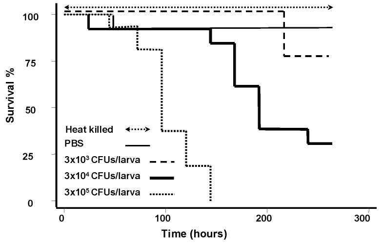
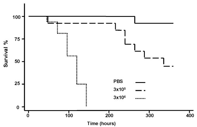
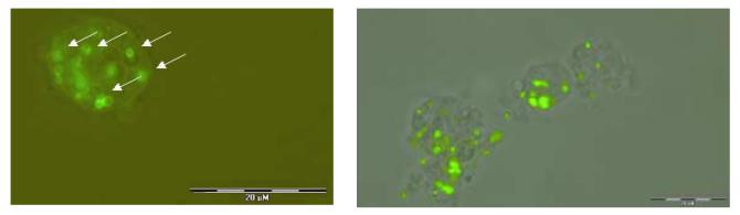
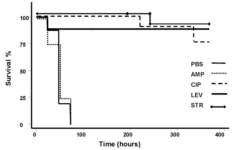
We used the killing of Galleria mellonella (Lepidoptera: Pyralidae; the greater wax moth) caterpillar by the live vaccine strain (LVS) of Francisella tularensis to develop an invertebrate host system that can be used to study F. tularensis infection and the in vivo effects of antibacterial compounds on F. tularensis LVS. After injection into the insect hemocoel, F. tularensis LVS, killed caterpillars despite the association of LVS with hemocytes. The rate of killing depended on the number of bacteria injected. Antibiotic therapy with ciprofloxacin, levofloxacin or streptomycin administered before or after inoculation prolonged survival and decreased the tissue burden of F. tularensis in the hemocoel. Delayed drug treatment reduced the efficacy of antibacterials and especially streptomycin. The G. mellonella-F. tularensis LVS system may facilitate the in vivo study of F. tularensis, efficacy with antibacterial agents.


Figures

Fig. 1
Fig. 1. Killing of G. mellonella caterpillars by F. tularensis LVS depends on the number of bacteria inoculated
Kaplan-Meier plots of G. mellonella survival after injection of different inocula of F. tularensis LVS. Injection with 3×10⁵ cfu/larva resulted in significantly higher death rate, compared to injection with 3×10⁴ cfu/larva (P<0.0001), or 3×10³ cfu/larva (P<0.0001). Injection with 3×10⁴ cfu/larva resulted in significantly higher death rate compared to injection with 3×10³ cfu/larva (P=0.0017). There was no killing of caterpillars that received heat killed bacterial cells of the same strain (3×10⁵ cells/larva). The caterpillars were maintained at 37°C.
Fig. 2
Fig. 2. Killing of G. mellonella caterpillars by F. tularensis LVS depends on the temperature after inoculation
Kaplan-Meier plots of G. mellonella survival after injection of different inocula of F. tularensis LVS. The caterpillars were maintained at 30°C. Injection with 3×10⁶ cfu/larva resulted in significantly higher death rate compared to controls and to injection with 3×10⁵ cfu/larvae. However, the killing rate at 30°C was slower than at 37°C (Figure 1), and at 30°C injection with 3×10⁵ cfu/larva was associated with significant survival (50% on Day 15).
Fig. 3
Fig. 3. Association of F. tularensis LVS cells with G. mellonella hemocytes
Hemocytes from caterpillars infected with F. tularensis LVS. After injection, caterpillars were maintained at 37°C and 48 hours after inoculation we collected their hemolymph. Following fixation, cells were diluted in Grace's insect medium and stained with mouse monoclonal antibody to F. tularensis lipopolysaccharide (LPS), as described under Materials and Methods. Left panel: Arrows point to FITC-labeled F. tularensis bacteria associated with a G. mellonella hemocyte. Right panel: Group of G. mellonella hemocytes with FITC-labeled associated F. tularensis bacteria.
Fig. 4
Fig. 4. Melanization is a part of the insect response to infection by F. tularensis LVS
Over the progress of the F. tularensis infection, caterpillars demonstrated obvious signs of progressive melanization. From right to left: G. mellonella at different time points after challenge with an inoculum of the LVS of F. tularensis 2 × 10⁶ cfu/larva. Infected larvae show progressively increasing melanization. From right to left larvae are at 0, 12, 24 (three larvae) and 48 hours (the last three larvae) after inoculation. Petri dish diameter is 8.5 cm.
Fig. 5
Fig. 5. Antibacterial agents prolong the survival of G. mellonella caterpillars after challenge with F. tularensis LVS
We examined the role of the most commonly used agents for F. tularensis infection by administering single doses of ciprofloxacin (CIP) 20 mg/kg, levofloxacin (LEV) 15 mg/kg, streptomycin (STR) 15 mg/kg or ampicillin (AMP) 150 mg/kg after the inoculation of caterpillars with 3×10⁶ cfu of F. tularensis LVS per larva. A control group received the F. tularensis inoculum and PBS instead of antibacterials. Treatment with ciprofloxacin, levofloxacin or streptomycin prolonged the survival of G. mellonella caterpillars (P< 0.0001 compared to control). However, ampicillin was not effective against F. tularensis LVS. The caterpillars were maintained at 37°C.
Figure 6
Figure 6. Antibacterial agents before challenge with F. tularensis LVS prolong the survival of G. mellonella caterpillars
A. We examined the role of the most commonly used agents for F. tularensis infection by administering a single dose of ciprofloxacin (CIP) 20 mg/kg, levofloxacin (LEV) 15 mg/kg, or streptomycin (STR) 15 mg/kg, one hour before infection with F. tularensis LVS 3×10⁶ cfu/larva. A control group received F. tularensis LVS and PBS instead of these drugs. Treatment with CIP, LEV or STR prolonged the survival of G. mellonella caterpillars (P<0.001 compared to control). Ampicillin (AMP) 150mg/kg given 1 hour before the infection with F. tularensis LVS 3×10⁶ cfu/larva did not protect G. mellonella caterpillars. B. We also examined the bacterial burden in the hemolymph after 48 hours using a single dose of ampicillin, ciprofloxacin, levofloxacin or streptomycin. A control group received treatment with PBS. CFU were counted per five larvae. The caterpillars were maintained at 37°C.

Similar articles

Cited by

References

    1. Tarnvik A. Nature of protective immunity to Francisella tularensis. Rev. Infect. Dis. 1989;11:440–451. - PubMed
    1. Prior RG, Klasson L, Larsson P, Williams K, Lindler L, Sjostedt A, Svensson T, Tamas I, Wren BW, Oyston PC, Andersson SG, Titball RW. Preliminary analysis and annotation of the partial genome sequence of Francisella tularensis strain Schu 4. J. Appl. Microbiol. 2001;91:614–620. - PubMed
    1. Dennis DT, Inglesby TV, Henderson DA, Bartlett JG, Ascher MS, Eitzen E, Fine AD, Friedlander AM, Hauer J, Layton M, Lillibridge SR, McDade JE, Osterholm MT, O'Toole T, Parker G, Perl TM, Russell PK, Tonat K. Tularemia as a biological weapon: medical and public health management. Jama. 2001;285:2763–2773. - PubMed
    1. Ellis J, Oyston PC, Green M, Titball RW. Tularemia. Clin. Microbiol. Rev. 2002;15:631–646. - PMC - PubMed
    1. Hernychova L, Stulik J, Halada P, Macela A, Kroca M, Johansson T, Malina M. Construction of a Francisella tularensis two-dimensional electrophoresis protein database. Proteomics. 2001;1:508–515. - PubMed

Publication types

LinkOut - more resources