Comparative Study
. 2007 Apr 10;104(15):6200-5.
doi: 10.1073/pnas.0609174104. Epub 2007 Mar 30.

Structural basis for PRYSPRY-mediated tripartite motif (TRIM) protein function

Affiliations
Comparative Study

Structural basis for PRYSPRY-mediated tripartite motif (TRIM) protein function

Leo C James et al. Proc Natl Acad Sci U S A. .

Abstract

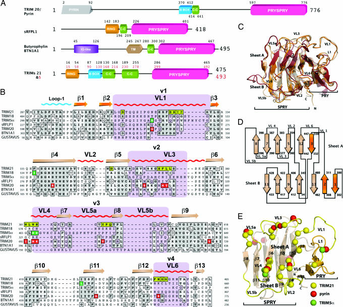
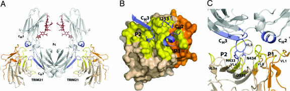
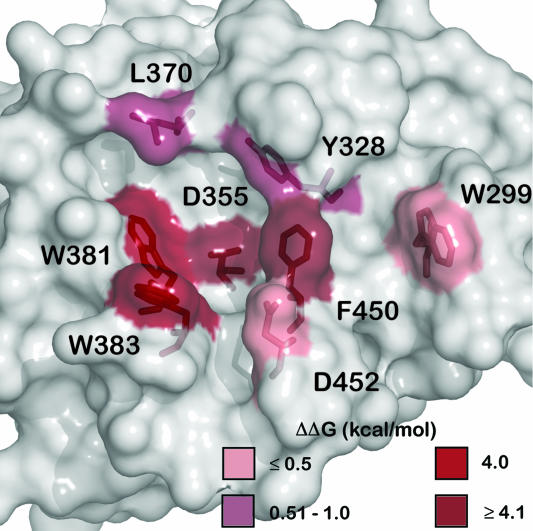
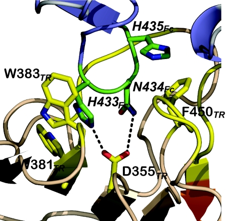
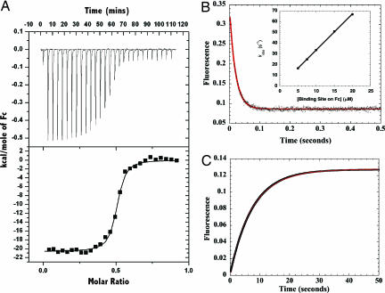
The human tripartite motif (TRIM) family comprises 70 members, including HIV restriction factor TRIM5alpha and disease-associated proteins TRIM20 (pyrin) and TRIM21. TRIM proteins have conserved domain architecture but diverse cellular roles. Here, we describe how the C-terminal PRYSPRY domain mediates diverse TRIM functions. The crystal structure of TRIM21 PRYSPRY in complex with its target IgG Fc reveals a canonical binding interface comprised of two discrete pockets formed by antibody-like variable loops. Alanine scanning of this interface has identified the hot-spot residues that control TRIM21 binding to Fc; the same hot-spots control HIV/murine leukemia virus restriction by TRIM5alpha and mediate severe familial Mediterranean fever in TRIM20/pyrin. Characterization of the IgG binding site for TRIM21 PRYSPRY reveals TRIM21 as a superantigen analogous to bacterial protein A and suggests that an antibody bipolar bridging mechanism may contribute to the pathogenic accumulation of anti-TRIM21 autoantibody immune complex in autoimmune disease.


Conflict of interest statement

The authors declare no conflict of interest.

Figures

Fig. 1.
Fig. 1.
The PRYSPRY domain has a conserved binding interface that dictates function in TRIM and other protein families. (A) Domain architecture of TRIM proteins 20, 21, and 5α, Butyrophylin BTN1A1, and putative protein sRFPL1. Domain boundaries as predicted by SMART are indicated (47). (B) Sequence alignment of TRIM21 PRYSPRY with those of disease-related TRIM proteins and homologous SPRY domains. TRIM21 secondary structure elements are indicated along with the position of the canonical binding loops (VLs). TRIM21:Fc contact residues are shown in yellow boxes. Dark green boxes indicate the position of mutations in TRIM5α. Red boxes indicate pyrin mutations that confer FMF susceptibility. The four variable regions known to dictate viral restriction specificity in TRIM5α are marked in purple. Gray shading indicates sequence similarity. (C) Superposition of TRIM21 (wheat), GUSTAVUS (red), and sRFPL1 (orange). (D) PRYSPRY topology cartoon. The PRY subdomain is colored in orange and the SPRY is colored in wheat. (E) TRIM21 PRYSPRY binding site. Variable binding loops are indicated in yellow. Yellow spheres correspond to important contacts with Fc, green indicates TRIM5α mutations, and red shows pyrin mutations linked to FMF.
Fig. 2.
Fig. 2.
The TRIM21 PRYSPRY:Fc complex. (A) Secondary structure representation of the complex. Contact areas on the Fc are marked in dark blue and on TRIM21 in yellow. The PRY subdomain of TRIM21 is marked in orange and SPRY in wheat. (B) Surface representation of the complex showing the two binding pockets (P1 and P2) and contacting Fc structure (blue). Yellow indicates surface buried upon complex formation. (C) Secondary structure representation of the TRIM21:Fc interface showing two discrete pockets and hydrogen-bond interactions between Fc residues H433 and N434 and TRIM21 D355.
Fig. 3.
Fig. 3.
Alanine scan of the TRIM21 PRYSPRY binding interface. Mutants that reduce affinity are indicated in red, the darker the color the greater the effect on the ΔΔG of binding.
Fig. 4.
Fig. 4.
TRIM21 PRYSPRY hot-spot residues bind Fc hot-spot residues. Secondary structure representation of the core binding interactions; TRIM21 (TR) is in orange with yellow side chains; Fc is in blue with green side chains.
Fig. 5.
Fig. 5.
Kinetic and thermodynamic analysis of the TRIM21 PRYSPRY domain binding to the IgG Fc region. (A) Titration of TRIM21 into IgG Fc measured by ITC shows that two molecules of TRIM21 bind to each IgG Fc with a Kd of 37 nM. (B) Rapid mixing of TRIM21 with IgG Fc in a stopped-flow experiment results in a fast monophasic fluorescence quench. The data are shown as black dots with the fit to a single exponential function shown as a solid red line. (Inset) A linear increase in kobs with increasing pseudofirst-order concentration of Fc indicates a single step binding mechanism. Linear regression yields kon = 3.5 × 10⁶ M−1·s−1. (C) Mixing of excess protein A outcompetes TRIM21 for binding to IgG Fc resulting in a fluorescence enhancement with an observed rate kdiss of 0.13 s−1 to yield a kinetic Kd (= kdiss/kon) of 37 nM.

References

    1. Nisole S, Stoye JP, Saib A. Nat Rev Microbiol. 2005;3:799–808. - PubMed
    1. Stremlau M, Perron M, Lee M, Li Y, Song B, Javanbakht H, Diaz-Griffero F, Anderson DJ, Sundquist WI, Sodroski J. Proc Natl Acad Sci USA. 2006;103:5514–5519. - PMC - PubMed
    1. Perron MJ, Stremlau M, Song B, Ulm W, Mulligan RC, Sodroski J. Proc Natl Acad Sci USA. 2004;101:11827–11832. - PMC - PubMed
    1. Song B, Gold B, O'Huigin C, Javanbakht H, Li X, Stremlau M, Winkler C, Dean M, Sodroski J. J Virol. 2005;79:6111–6121. - PMC - PubMed
    1. Yap MW, Nisole S, Stoye JP. Curr Biol. 2005;15:73–78. - PubMed

Publication types

MeSH terms

Associated data

LinkOut - more resources