. 2007 May;73(10):3144-50.
doi: 10.1128/AEM.02937-06. Epub 2007 Mar 30.

Shiga toxin gene loss and transfer in vitro and in vivo during enterohemorrhagic Escherichia coli O26 infection in humans

Affiliations

Shiga toxin gene loss and transfer in vitro and in vivo during enterohemorrhagic Escherichia coli O26 infection in humans

Martina Bielaszewska et al. Appl Environ Microbiol. 2007 May.

Abstract

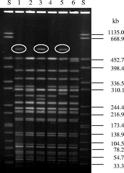
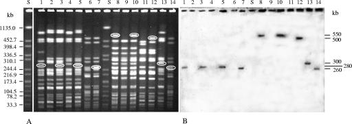
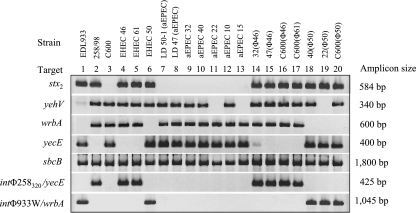
Escherichia coli serogroup O26 consists of enterohemorrhagic E. coli (EHEC) and atypical enteropathogenic E. coli (aEPEC). The former produces Shiga toxins (Stx), major determinants of EHEC pathogenicity, encoded by bacteriophages; the latter is Stx negative. We have isolated EHEC O26 from patient stools early in illness and aEPEC O26 from stools later in illness, and vice versa. Intrapatient EHEC and aEPEC isolates had quite similar pulsed-field gel electrophoresis (PFGE) patterns, suggesting that they might have arisen by conversion between the EHEC and aEPEC pathotypes during infection. To test this hypothesis, we asked whether EHEC O26 can lose stx genes and whether aEPEC O26 can be lysogenized with Stx-encoding phages from EHEC O26 in vitro. The stx2 loss associated with the loss of Stx2-encoding phages occurred in 10% to 14% of colonies tested. Conversely, Stx2- and, to a lesser extent, Stx1-encoding bacteriophages from EHEC O26 lysogenized aEPEC O26 isolates, converting them to EHEC strains. In the lysogens and EHEC O26 donors, Stx2-converting bacteriophages integrated in yecE or wrbA. The loss and gain of Stx-converting bacteriophages diversifies PFGE patterns; this parallels findings of similar but not identical PFGE patterns in the intrapatient EHEC and aEPEC O26 isolates. EHEC O26 and aEPEC O26 thus exist as a dynamic system whose members undergo ephemeral interconversions via loss and gain of Stx-encoding phages to yield different pathotypes. The suggested occurrence of this process in the human intestine has diagnostic, clinical, epidemiological, and evolutionary implications.


Figures

FIG. 1.
FIG. 1.
XbaI-digested genomic DNA from EHEC and aEPEC O26:H11 strains isolated from initial and follow-up stools, respectively, of three patients during a HUS outbreak. Lane 1, EHEC O26 (strain 50), patient A; lane 2, aEPEC O26 (strain 40), patient A; lane 3, EHEC O26 (strain 140), patient B; lane 4, aEPEC O26 (strain 41), patient B; lane 5, EHEC O26 (strain 141), patient C; lane 6, aEPEC O26 (strain 42), patient C; lanes S, molecular size standards (S. enterica serovar Braenderup strain H9812; Centers for Disease Control and Prevention, Atlanta, GA). XbaI fragments containing stx2 as demonstrated by hybridization with an stxA2 probe are circled.
FIG. 2.
FIG. 2.
PFGE (A) and stxA2 hybridization (B) of XbaI-digested genomic DNA from EHEC O26 phage donors, aEPEC O26 recipients, and lysogens transduced with stx2-harboring bacteriophages from EHEC O26. Lanes 1, EHEC O26 strain 46 (donor of phage φ46); lanes 2, aEPEC O26 strain 47; lanes 3, lysogen 47(φ46); lanes 4, aEPEC O26 strain 32; lanes 5, lysogen 32(φ46); lanes 6, E. coli strain C600; lanes 7, lysogen C600(φ46); lanes 8, EHEC O26 strain 50 (donor of phage φ50); lanes 9, aEPEC O26 strain 40; lanes 10, lysogen 40(φ50); lanes 11, aEPEC O26 strain 22; lanes 12, lysogen 22(φ50); lanes 13, EHEC O26 strain 61 (donor of phage φ61); lanes 14, lysogen C600(φ61); lanes S, molecular size standards (S. enterica serovar Braenderup strain H9812; Centers for Disease Control and Prevention, Atlanta, GA). The XbaI fragments that hybridized with the stxA2 probe are circled in panel A, and their sizes are given in panel B.
FIG. 3.
FIG. 3.
PCR analyses of phage integration sites in EHEC O26, aEPEC O26, and lysogens. Strains tested, loci examined, and lengths of resulting amplicons are listed across the top and to the left and right of the rows of amplicons, respectively. stx2-negative laboratory derivatives (LD) 47 and 50-1 fit the definition of aEPEC. Strains EDL933 (stx1- and stx2-harboring phages integrated in yehV and wrbA, respectively) (35, 37), 258/98 (stx2-harboring phage φ258320 integrated in yecE) (7), and E. coli K-12 C600 (all the genes investigated as putative phage integration sites intact) (8) were used as controls. In PCRs targeting yehV, wrbA, yecE, and sbcB, the presence of an amplicon indicates that the locus is intact, whereas the absence of an amplicon (or a very weak amplicon) indicates that the locus is occupied by foreign DNA. In PCRs connecting yecE with the int gene of phage φ258320 and wrbA with the int gene of phage φ933W (rows 6 and 7, respectively), the presence of an amplicon indicates that a phage with a homologous int gene is integrated in the respective locus; the absence of an amplicon indicates the absence of such a phage.

References

    1. Acheson, D. W. K., J. Reidl, X. Zhang, G. T. Keusch, J. J. Mekalanos, and M. K. Waldor. 1998. In vivo transduction with Shiga toxin 1-encoding phage. Infect. Immun. 66:4496-4498. - PMC - PubMed
    1. Allerberger, F., A. W. Friedrich, K. Grif, M. P. Dierich, H. J. Dornbusch, C. J. Mache, E. Nachbaur, M. Freilinger, P. Rieck, M. Wagner, A. Caprioli, H. Karch, and L. B. Zimmerhackl. 2003. Hemolytic-uremic syndrome associated with enterohemorrhagic Escherichia coli O26:H− infection and consumption of unpasteurized cow's milk. Int. J. Infect. Dis. 7:42-45. - PubMed
    1. Anjum, M. F., S. Lucchini, A. Thompson, J. C. Hinton, and M. J. Woodward. 2003. Comparative genomic indexing reveals the phylogenomics of Escherichia coli pathogens. Infect. Immun. 71:4674-4683. - PMC - PubMed
    1. Barton, C., L.-K. Ng, S. D. Tyler, and C. G. Clark. 2007. Temperate bacteriophages affect pulsed-field gel electrophoresis patterns of Campylobacter jejuni. J. Clin. Microbiol. 45:386-391. - PMC - PubMed
    1. Besser, T. E., N. Shaikh, N. J. Holt, P. I. Tarr, M. E. Konkel, P. Malik-Kale, C. W. Walsh, T. Whittam, and J. L. Bono. 2007. Greater diversity of Shiga toxin-encoding bacteriophage insertion sites among Escherichia coli O157:H7 isolates from cattle than in those from humans. Appl. Environ. Microbiol. 73:671-679. - PMC - PubMed

Publication types

MeSH terms