. 2007 May;6(5):855-67.
doi: 10.1128/EC.00399-06. Epub 2007 Mar 30.

Chitosan, the deacetylated form of chitin, is necessary for cell wall integrity in Cryptococcus neoformans

Affiliations

Chitosan, the deacetylated form of chitin, is necessary for cell wall integrity in Cryptococcus neoformans

Lorina G Baker et al. Eukaryot Cell. 2007 May.

Abstract

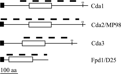
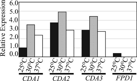
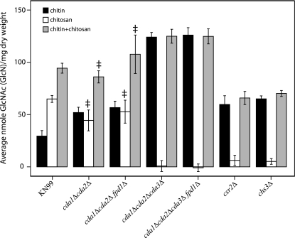
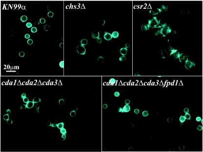
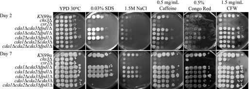
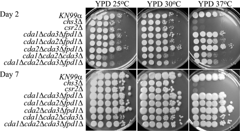
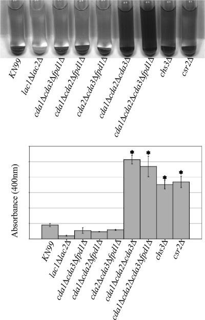
Cryptococcus neoformans is an opportunistic fungal pathogen that causes cryptococcal meningoencephalitis, particularly in immunocompromised patients. The fungal cell wall is an excellent target for antifungal therapies as it is an essential organelle that provides cell structure and integrity, it is needed for the localization or attachment of known virulence factors, including the polysaccharide capsule, melanin, and phospholipase, and it is critical for host-pathogen interactions. In C. neoformans, chitosan produced by the enzymatic removal of acetyl groups from nascent chitin polymers has been implicated as an important component of the vegetative cell wall. In this study, we identify four putative chitin/polysaccharide deacetylases in C. neoformans. We have demonstrated that three of these deacetylases, Cda1, Cda2, and Cda3, can account for all of the chitosan produced during vegetative growth in culture, but the function for one, Fpd1, remains undetermined. The data suggest a model for chitosan production in vegetatively growing C. neoformans where the three chitin deacetylases convert chitin generated by the chitin synthase Chs3 into chitosan. Utilizing a collection of chitin/polysaccharide deacetylase deletion strains, we determined that during vegetative growth, chitosan helps to maintain cell integrity and aids in bud separation. Additionally, chitosan is necessary for maintaining normal capsule width and the lack of chitosan results in a "leaky melanin" phenotype. Our analysis indicates that chitin deacetylases and the chitosan made by them may prove to be excellent antifungal targets.


Figures

FIG. 1.
FIG. 1.
Predicted protein structure of C. neoformans deacetylases. The name of each protein is indicated to the right of the protein structure. Domains were identified by BLAST searches of the NCBI database (www.ncbi.nlm.nih.gov/structure/cdd/wrpsb.cgi). The N-terminal signal sequence and GPI anchor were determined. Domains are identified as follows: black rectangles, N-terminal signal sequence; white rectangles, polysaccharide deacetylase domain; omega, predicted GPI cleavage site. The dashed line above each protein indicates the extent of deletion in the corresponding genomic region for each deacetylase gene. aa, amino acids.
FIG. 2.
FIG. 2.
Expression analysis of deacetylases during vegetative growth of C. neoformans. Liquid cultures of KN99α were grown in YPD for 24 h at 25°C, 30°C, and 37°C. Native deacetylase expression was normalized to actin expression, and relative expression values were determined by ImageQuant (version 5.1). Gene-specific probe and growth conditions are listed beneath the graph. The graph depicts one of three independent biological replicates (each having similar results).
FIG. 3.
FIG. 3.
Eosin Y staining of chitosan in fungal cells. C. neoformans cells were grown for 24 h, S. cerevisiae cells were grown for 48 h, and all cells were then stained with eosin Y. Bright field and fluorescein isothiocyanate panels are shown for each strain. Strains or deletions are indicated at the top left of panels, and all cells were grown vegetatively except for those in the top right panel depicting sporulating S. cerevisiae. Arrows indicate cells that have taken up eosin Y, and these cells are dead, as confirmed by trypan blue staining (data not shown). All photos are the same magnification (×1,000).
FIG. 4.
FIG. 4.
Chitin and chitosan contents of C. neoformans KN99α and selected deletion strains. Measurements of strains were taken at 70 h of growth in liquid YPD at 25°C and reported as average nanomoles per milligram of dry weight of chitin, chitosan, and the total chitin plus chitosan as indicated. Each measurement represents the average of three or more independent cultures for each strain. Double daggers indicate measurements that were not significantly different from that of the wild type (P > 0.001). Strains or deletions are indicated at the bottom of the graph. Error bars indicate standard deviations.
FIG. 5.
FIG. 5.
CFW staining of C. neoformans KN99α and deletion strains. Cells were grown in YPD at 30°C and stained with CFW, a dye that binds to chitin. Strains or deletions are indicated top left of each frame. Scale bar = 20 μm. All photos are at the same magnification (×1,000).
FIG. 6.
FIG. 6.
Analysis of KN99α and deletion strains for in vitro sensitivity to cell wall stressors. Strains or deletion mutants were grown in YPD medium to mid-log phase, serially diluted 10-fold (starting with 1 × 10⁵ cells in the left column of each panel), and plated on YPD solid medium and YPD solid medium supplemented with various cell wall stressors. The times of incubation are indicated at the far left. Strains are indicated at the left of the panels. Incubation conditions are indicated at the top of the panels. All plates containing cell wall stressors were incubated at 30°C.
FIG. 7.
FIG. 7.
Analysis of KN99α and deletion strains for in vitro sensitivity to temperature. The experiment was carried out as described in the legend for Fig. 6.
FIG. 8.
FIG. 8.
Melanin production of chitin synthesis and deacetylation deletion strains. (Top panel) Cells were grown 2 days on YPD at 30°C, resuspended to identical optical densities in glucose-free asparagine medium containing l-DOPA, and allowed to shake at 30°C. Cells were centrifuged to visualize the secreted pigment and the color of the pellets. Pictures were taken at 24 h. The strain or deletion is indicated at the bottom of the panel. The photo is representative of four independent experiments. A lac1Δlac2Δ strain is included as a negative control. (Bottom panel). Quantification of the OD400 of the supernatants from the cultures as shown in the top panel. Asterisk indicates significant differences between supernatant at OD400 compared to that of the wild type by using a two-tailed equal-variance Student t test. P < 0.001. Error bars indicate standard deviations.
FIG. 9.
FIG. 9.
India ink staining of capsule of KN99α and deletion strains. Strains were grown for 5 days on DME medium at 30°C. Strains or deletion mutants are indicated at top left of each frame. Cells were mixed with a 1:4 dilution of India ink-to-H2O solution. Capsule widths of more than 200 cells/strain were measured from the edge of the cell wall to the distal edge of the capsule, and average capsule width is indicated on the bottom right of each frame. A cap59Δ strain is included as a negative control. Scale bar = 20 μm.

Similar articles

Cited by

References

    1. Adams, D. J. 2004. Fungal cell wall chitinases and glucanases. Microbiology 150:2029-2035. - PubMed
    1. Banks, I. R., C. A. Specht, M. J. Donlin, K. J. Gerik, S. M. Levitz, and J. K. Lodge. 2005. A chitin synthase and its regulator protein are critical for chitosan production and growth of the fungal pathogen Cryptococcus neoformans. Eukaryot. Cell 4:1902-1912. - PMC - PubMed
    1. Biondo, C., C. Beninati, M. Bombaci, L. Messina, G. Mancuso, A. Midiri, R. Galbo, and G. Teti. 2003. Induction of T helper type 1 responses by a polysaccharide deacetylase from Cryptococcus neoformans. Infect. Immun. 71:5412-5417. - PMC - PubMed
    1. Biondo, C., C. Beninati, D. Delfino, M. Oggioni, G. Mancuso, A. Midiri, M. Bombaci, G. Tomaselli, and G. Teti. 2002. Identification and cloning of a cryptococcal deacetylase that produces protective immune responses. Infect. Immun. 70:2383-2391. - PMC - PubMed
    1. Bose, I., A. J. Reese, J. J. Ory, G. Janbon, and T. L. Doering. 2003. A yeast under cover: the capsule of Cryptococcus neoformans. Eukaryot. Cell 2:655-663. - PMC - PubMed

Publication types

MeSH terms