. 2007 Mar;19(3):750-66.
doi: 10.1105/tpc.107.050385. Epub 2007 Mar 30.

Elaboration of B gene function to include the identity of novel floral organs in the lower eudicot Aquilegia

Affiliations

Elaboration of B gene function to include the identity of novel floral organs in the lower eudicot Aquilegia

Elena M Kramer et al. Plant Cell. 2007 Mar.

Abstract

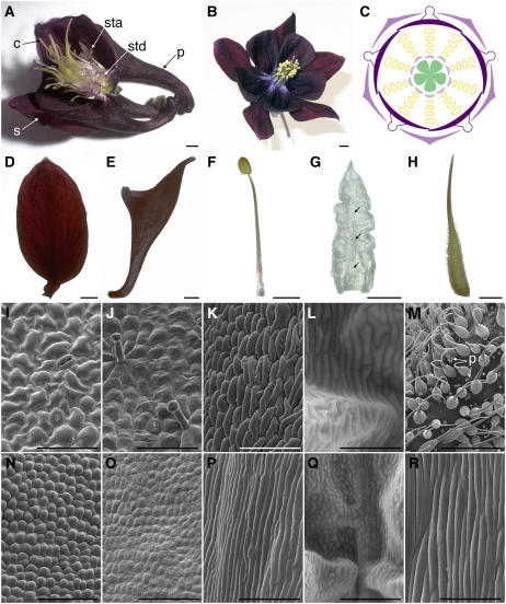
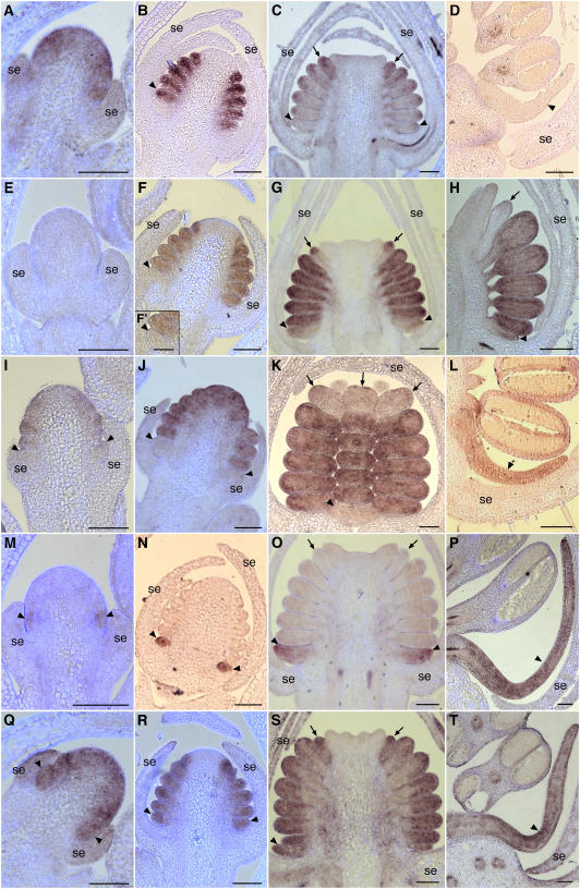
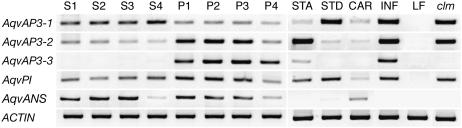
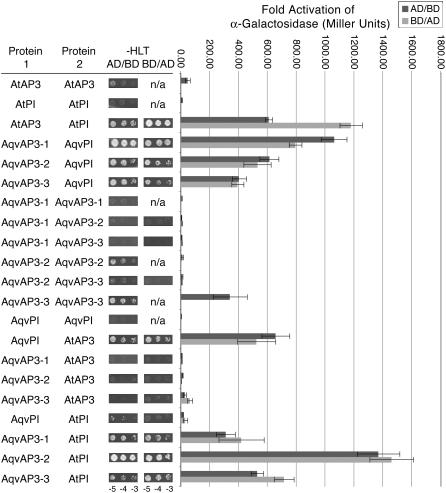
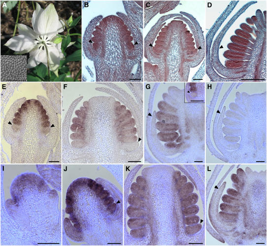
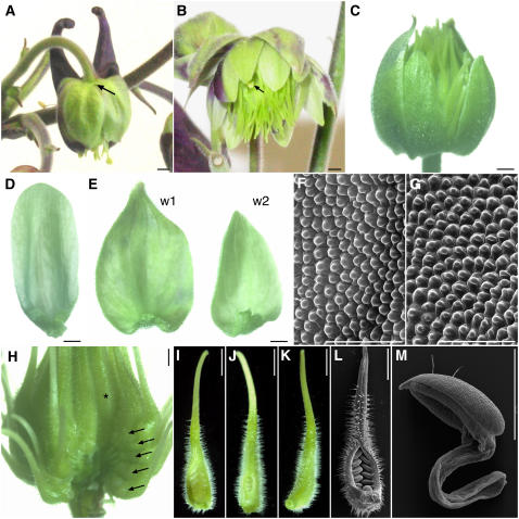
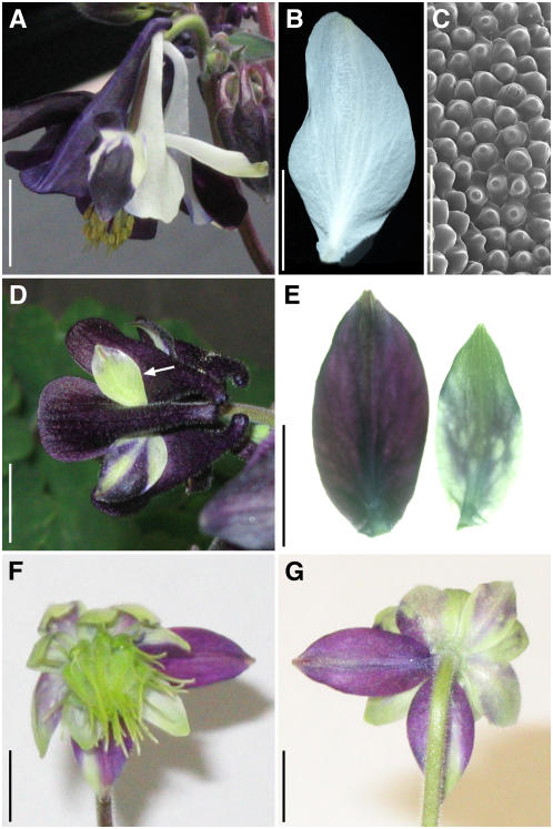
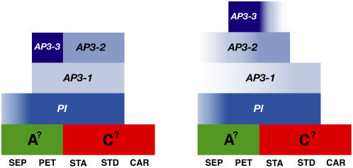
The basal eudicot Aquilegia (columbine) has an unusual floral structure that includes two morphologically distinct whorls of petaloid organs and a clearly differentiated fifth organ type, the staminodium. In this study, we have sought to determine how Aquilegia homologs of the B class genes APETALA3 (AP3) and PISTILLATA (PI) contribute to these novel forms of organ identity. Detailed expression analyses of the three AP3 paralogs and one PI homolog in wild-type and floral homeotic mutant lines reveal complex patterns that suggest that canonical B class function has been elaborated in Aquilegia. Yeast two-hybrid studies demonstrate that the protein products of Aquilegia's AP3 and PI homologs can form heterodimers, much like what has been observed for their core eudicot homologs. Downregulation of AqvPI using virus-induced gene silencing indicates that in addition to petal and stamen identity, this locus is essential to staminodial identity but may not control the identity of the petaloid sepals. Our findings show that preexisting floral organ identity programs can be partitioned and modified to produce additional organ types. In addition, they indicate that some types of petaloid organs are not entirely dependent on AP3/PI homologs for their identity.


Figures

Figure 1.
Figure 1.
Floral Morphology of Wild-Type A. vulgaris. (A), (B), and (D) to (H) Light micrographs. (I) to (K), (M) to (P), and (R) Scanning electron micrographs. (L) and (Q) Environmental scanning electron micrographs. (A) Side view of a flower at anthesis. Two sepals, one petal, and several whorls of stamens were removed to expose the staminodia. c, carpel; p, petal; s, sepal; sta, stamen; std, staminodium. (B) Head-on view of a flower at anthesis. (C) Floral diagram showing relative positions of five organ types. Whorls one and two are composed of 5 sepals and 5 petals, respectively, followed by four to seven whorls of 10 stamens, one whorl of 10 staminodia, and an innermost whorl of 5 free carpels. (D) Sepal at anthesis. (E) Petal at anthesis. (F) Stamen close to anthesis. (G) Staminodium at anthesis. The midvein of the staminodium is marked by arrows. (H) Carpel at anthesis. (I) and (N) Scanning electron microscopy images of the abaxial (I) and adaxial (N) surfaces of the sepal. (J) and (O) Scanning electron microscopy images of the abaxial (J) and adaxial (O) surfaces of the petal. (K) and (P) Scanning electron microscopy images of the anther (K) and filament (P) of the stamen. (L) and (Q) Environmental scanning electron microscopy images of the staminodium. (M) and (R) Scanning electron microscopy images of the ovary wall (M) and style (R) of the carpel. Small spheres in (M) are pollen (p). Bars = 2 mm in (A), (B), (F), and (G), 2.5 mm in (D), (E), and (H), 50 μm in (I), (J), and (L), 100 μm in (K) and (M) to (P), 250 μm in (Q), and 300 μm in (R).
Figure 2.
Figure 2.
In Situ Hybridization of AqvAP3-1, AqvAP3-2, AqvAP3-3, and AqvPI in Wild-Type Floral Meristems. (A) to (D) AqvAP3-1. (E) to (L) AqvAP3-2. (M) to (P) AqvAP3-3. (Q) to (T) AqvPI. (A), (E), and (M) show very early-stage floral meristems in which the sepals are just emerging. (I) and (Q) are slightly later, as the petal and stamen primordia are just beginning to initiate. (B), (F), (J), (N), and (R) show an early-stage floral meristem in which the sepals are growing to enclose the floral meristem and the petal, stamen, and staminodium primordia are initiating. (F′) is a magnification of the petal primordium shown in (F). (C), (G), (K), (O), and (S) correspond to the stage at which the carpel primordia are appearing and the petals, stamens, and staminodia are beginning to become morphologically distinct. (D), (H), (L), (P), and (T) show a range of later stages in which the various floral organ types are differentiating. The petals are elongating and beginning to become spurred, microsporogenesis is under way in the stamens, and the staminodia are clearly differentiated from the fertile stamens. (D), (J), (K), and (T) are tangential longitudinal sections, while the others are all radial longitudinal. se, sepals; arrowheads indicate petal primordia, and arrows indicate staminodia. The asterisk in (P) indicates weak AqvAP3-3 expression in the connective. All primordia located between the petals and staminodia are fertile stamens. Bars = 100 μm.
Figure 3.
Figure 3.
Locus-Specific RT-PCR of RNA Prepared from Organs Corresponding to Stages S1 to S4 and P1 to P4 as Shown in Supplemental Figures 1 and 2 Online. Additional reactions were performed using RNA from stamens (STA), staminodia (STD), carpels (CAR), wild-type inflorescences (INF), prevernalization vegetative leaves (LF), and clm mutant inflorescences. Six genes were analyzed: AqvAP3-1, AqvAP3-2, AqvAP3-3, AqvPI, AqvANS, and ACTIN.
Figure 4.
Figure 4.
Protein Interactions between AP3 and PI Homologs of A. vulgaris as Determined by Growth on Selective SD Medium and Quantification of Secreted α-Galactosidase Activity (see Supplemental Methods Online). Aquilegia and Arabidopsis proteins were cloned into both binding domain (BD) and activation domain (AD) constructs. For each protein pair listed, AD/BD indicates that protein 1 was fused with AD and protein 2 was fused with BD. BD/AD indicates the opposite situation. The colonies shown in the –HLT (−HisLeuTrp) columns represent dilution series (105, 104, and 103 colony-forming units) of each strain grown on –HLT SD medium supplemented with 20 mM 3-amino-1,2,4-triazole. n/a, not applicable. For the graph, the dark gray bars show the results from analyses of α-galactosidase activity where the first protein listed was in the AD construct and the second protein was in the BD construct. The light gray bars are for the reverse BD/AD combination. Error bars represent sd across three independent replicates.
Figure 5.
Figure 5.
Floral Morphology of A. vulgaris clm and in Situ Hybridization of AqvAP3-1, AqvAP3-2, AqvAP3-3, and AqvPI in clm Floral Meristems. (A) Photograph of a flower from the underside. All perianth organs have the morphology of sepals (inset shows adaxial epidermis). (B) to (D) Histological sections of developing floral meristems. Arrowheads indicate second whorl organs. (E) and (F) AqvAP3-1. (G) AqvAP3-2 with inset showing earlier stage. (H) AqvAP3-3. (I) to (L) AqvPI. Sections in (B), (C), and (E) are tangential longitudinal, while those in (D) and (F) to (L) are radial longitudinal. Arrowheads indicate second whorl primordia, unless stated otherwise. Bars = 100 μm.
Figure 6.
Figure 6.
Phenotypes of AqvPI-AqvANS–Silenced Floral Buds and Organs. (A) Flower showing a moderate silencing phenotype. Half of the perianth shows AqvANS silencing, and the petal spur on that side of the flower is missing (arrow indicates the expected position). (B) Flower showing a strong silencing phenotype. The perianth organs show only patchy AqvANS expression, all of the second whorl organs have the appearance of sepals, and the majority of internal organs are transformed into carpeloid organs. One stamen-like organ is indicated by an arrow. (C) Flower showing a strong silencing phenotype. Two sepals were removed to expose the inner organs. No AqvANS expression is apparent, the second whorl organs are completely transformed into sepals, and all internal organs are carpels. (D) A silenced second whorl organ from the flower shown in (A). (E) Organs from the first (w1) and second (w2) whorls of the flower shown in (C). (F) Scanning electron microscopy of the adaxial epidermal surface of a silenced second whorl organ. (G) Scanning electron microscopy of the adaxial epidermal surface of a silenced first whorl organ. (H) The carpeloid organs of a strongly silenced flower. The perianth as well as one vertical orthostichy of carpeloid organs has been removed. Arrows indicate a vertical row of transformed organs, which show carpel identity in every whorl through to the innermost whorl (asterisk), representing the position of the wild-type carpels. (I) to (K) Individual carpeloid organs from strongly silenced flowers exhibiting a range of partial fusion. (L) Scanning electron microscopy image of the organ shown in (I). (M) Scanning electron microscopy image of a staminoid organ from a strongly silenced flower. Bars = 1 mm in (A) to (E) and (H) to (M) and 100 μm in (F) and (G).
Figure 7.
Figure 7.
Locus-Specific RT-PCR on RNA Prepared from Organs of VIGS-Treated Flowers. (A) RNA samples from five tissue types were tested: flowers that were treated with TRV2-AqvPI-AqvANS but did not show silencing (NS), silenced flowers that were treated with TRV2-AqvANS (ANS−), first whorl organs from strongly silenced flowers treated with TRV2-AqvPI-AqvANS (w1), second whorl organs from strongly silenced flowers treated with TRV2-AqvPI-AqvANS (w2), and carpeloid organs from strongly silenced flowers treated with TRV2-AqvPI-AqvANS (car). (B) Individual RT-PCR for 6 unsilenced and 12 silenced tissue samples. The sample numbers correspond to the bud number and whorl from which the sample was taken. For instance, sample 2.2 was a second whorl organ from the second flower to be characterized. We analyzed 33 individual flowers showing some degree of silencing as well as 12 flowers showing no silencing from the same plants. In some cases, silenced and unsilenced organs from the same whorl were analyzed separately (e.g., 3.1a and 3.1b). Unsilenced samples included sepals (whorl 1), petals (whorl 2), and stamens (whorl 3), while silenced samples included sepals (whorl 1), petaloid or sepaloid organs in whorl 2, and carpeloid organs in internal whorls (c). Normalized expression values are shown for each sample.
Figure 8.
Figure 8.
Sepal Phenotypes in Flowers Treated with TRV2-AqvANS or TRV2-AqvPI-AqvANS. (A) A flower showing silencing in response to treatment with TRV2-AqvANS. (B) A single AqvANS-silenced sepal. (C) Scanning electron microscopy image of the adaxial epidermis of an AqvANS-silenced sepal. (D) A flower showing patchy silencing in response to treatment with TRV2-AqvPI-AqvANS. Note the small, green sepal (arrow). (E) Sepals from the same whorl of a single flower with partial AqvPI-AqvANS silencing. Note the size difference between the unsilenced (left) and silenced (right) organs. (F) and (G) Front (F) and back (G) views of a flower showing strong AqvPI-AqvANS silencing. Two first whorl organs are relatively unsilenced and show distinct expansion patterns relative to the more strongly silenced organs. Bars = 10 mm in (A), (B), and (D) to (G) and 100 μm in (C).
Figure 9.
Figure 9.
Modified ABC Models Based on the Early (Left) and Late (Right) Expression Patterns of AP3 and PI Homologs in Aquilegia. The early-stage model corresponds to the period when sepals, stamens, and staminodia are initiating, while the late-stage model encompasses everything after carpel initiation. Color gradients indicate transient or weak expression. Each AP3 paralog has a distinct expression pattern, while the expression domain of the single PI homolog encompasses the summation of all of the AP3 expression domains. Placeholders are included for potential A and C function loci. SEP, sepals; PET, petals; STA, stamens; STD, staminodia; CAR, carpels.

Similar articles

Cited by

References

    1. Aagaard, J.E., Olmstead, R.G., Willis, J.H., and Phillips, P.C. (2005). Duplication of floral regulatory genes in the Lamiales. Am. J. Bot. 92 1284–1293. - PubMed
    1. Albert, V.A., Gustafsson, M.H.G., and Di Laurenzio, L. (1998). Ontogenetic systematics, molecular developmental genetics, and the angiosperm petal. In Molecular Systematics of Plants, Vol. II, D. Soltis, P. Soltis, and J.J. Doyle, eds (New York: Chapman and Hall), pp. 349–374.
    1. Ambrose, B.A., Lerner, D.R., Ciceri, P., Padilla, C.M., Yanofsky, M.F., and Schmidt, R.J. (2000). Molecular and genetic analyses of the Silky1 gene reveal conservation in floral organ specification between eudicots and monocots. Mol. Cell 5 569–579. - PubMed
    1. Aoki, S., Uehara, K., Imafuku, M., Hasebe, M., and Ito, M. (2004). Phylogeny and divergence of basal angiosperms inferred from APETALA3- and PISTILLATA-like MADS-box genes. J. Plant Res. 117 229–244. - PubMed
    1. Becker, A., and Theissen, G. (2003). The major clades of MADS-box genes and their role in the development and evolution of flowering plants. Mol. Phylogenet. Evol. 29 464–489. - PubMed

Publication types

MeSH terms

Associated data