. 2007 Mar;19(3):847-61.
doi: 10.1105/tpc.106.044123. Epub 2007 Mar 30.

Rice UDP-glucose pyrophosphorylase1 is essential for pollen callose deposition and its cosuppression results in a new type of thermosensitive genic male sterility

Affiliations

Rice UDP-glucose pyrophosphorylase1 is essential for pollen callose deposition and its cosuppression results in a new type of thermosensitive genic male sterility

Rongzhi Chen et al. Plant Cell. 2007 Mar.

Abstract

UDP-glucose pyrophosphorylase (UGPase) catalyzes the reversible production of glucose-1-phosphate and UTP to UDP-glucose and pyrophosphate. The rice (Oryza sativa) genome contains two homologous UGPase genes, Ugp1 and Ugp2. We report a functional characterization of rice Ugp1, which is expressed throughout the plant, with highest expression in florets, especially in pollen during anther development. Ugp1 silencing by RNA interference or cosuppression results in male sterility. Expressing a double-stranded RNA interference construct in Ugp1-RI plants resulted in complete suppression of both Ugp1 and Ugp2, together with various pleiotropic developmental abnormalities, suggesting that UGPase plays critical roles in plant growth and development. More importantly, Ugp1-cosuppressing plants contained unprocessed intron-containing primary transcripts derived from transcription of the overexpression construct. These aberrant transcripts undergo temperature-sensitive splicing in florets, leading to a novel thermosensitive genic male sterility. Pollen mother cells (PMCs) of Ugp1-silenced plants appeared normal before meiosis, but during meiosis, normal callose deposition was disrupted. Consequently, the PMCs began to degenerate at the early meiosis stage, eventually resulting in complete pollen collapse. In addition, the degeneration of the tapetum and middle layer was inhibited. These results demonstrate that rice Ugp1 is required for callose deposition during PMC meiosis and bridges the apoplastic unloading pathway and pollen development.


Figures
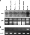
Figure 1.
Figure 1.
Expression Patterns of Ugp1 and Ugp2 in Wild-Type Hejiang 19 Rice Plants. (A) RNA gel blot analysis of Ugp1 transcript levels in wild-type Hejiang 19 rice plants. Total RNA (20 μg) extracted from wild-type tissues, including roots, seedling stems, mature stems, seedling leaves, mature leaves, and pooled florets at various stages before flowering was probed with full-length Ugp1 cDNA. An ethidium bromide stain of the gel is shown to confirm equal RNA loading. (B) RT-PCR analysis of Ugp2 expression in wild-type Hejiang 19 rice plants. The amplification of the rice Actin1 gene was used as a control to show that approximately equal amounts of total RNA had been used in the RT-PCR analysis.
Figure 2.
Figure 2.
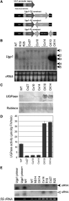
Ugp1 Expression Patterns in Transgenic Rice Plants. (A) Structures of constructs for rice transformation. The promoter of the maize ubiquitin 1 gene of ∼2 kb comprises, in the 5′ to 3′ direction, the following: a promoter with a transcription start site (+1); two overlapping heat shock elements located at positions −214 and −204 from the transcription start site; an 83-bp leader sequence adjacent to the transcription start site (exon); an intron of ∼1 kb; and a translation start site. The overexpression construct (Ugp1-OX), antisense construct (Ugp1-AS), and dsRNAi construct (Ugp1-RI) of Ugp1 were developed under the control of the Ubi1 promoter and nopaline synthase (Nos) terminator cassette. Arrows represent the primers used to generate the RT-PCR products shown in Figure 7D. (B) RNA gel blot analysis of Ugp1 transcript levels in transgenic rice. Total RNA extracted from rice plants at the heading stage was probed with full-length Ugp1 cDNA. Arrowheads indicate (1) the unprocessed longer-than-full-length transcript and (2) endogenous Ugp1 mRNA; (3) to (5) indicate the silencing-related RNA degradation intermediates. Loading of equal amounts of RNA was confirmed by ethidium bromide staining. (C) Protein gel blot analysis of UGPase protein from transgenic plants using an antibody against rice UGPase. Equal loading of proteins in each lane was confirmed by probing a duplicate blot with an anti-ribulose-1,5-bisphosphate carboxylase/oxygenase (Rubisco) antibody. (D) UGPase activities in transgenic plants. UGPase activities are shown as means ± se (n = 3). (E) siRNA analysis in transgenic plants. Seventy-five nanograms of Ugp1 primers (18 and 21 nucleotides) were intermixed or mixed with wild-type RNA and served as size standard and hybridization controls. Closed arrowhead indicates the siRNA probed by the Ugp1 cDNA fragment between +333 and +852 (related to the ATG at +1 bp), and the open arrowhead indicates the siRNA by the 3′-end of the Ugp1 coding region. 5S rRNA was used as the loading control. Co10, Co14, and Co27, cosuppressing plants of lines 10, 14, and 27, respectively; OX10 and OX14, overexpressing plants of lines 10 and 14, respectively; NS14, null segregant plant of line 14; R19 and R25, RNAi plants of lines 19 and 25, respectively.
Figure 3.
Figure 3.
Phenotypes of Ugp1-Silenced Plants. (A) Wild-type plant at maturity stage. (B) Cosuppressing plant at maturity stage showing no seed set on the panicles. (C) RNAi plant at maturity stage. (D) Wild-type spikelets at the heading stage. (E) Cosuppressing plant spikelets at the heading stage. (F) Flower and anther morphology of a wild-type plant. (G) Flower and anther morphology of a cosuppressing plant. (H) Wild-type pollen grains stained with KI-I2 solution. (I) Cosuppressing plant pollen grains stained with KI-I2 solution. Co27, cosuppressing plant of line 27; R19, RNAi plant of line 19. Bar = 10 cm.
Figure 4.
Figure 4.
Anther Development in Wild-Type Plants and Cosuppressing Plants. The micrographs show one of the four lobes in cross sections of wild-type anthers ([A] to [G]) and anthers from cosuppressing plants ([H] to [N]) at the premeiosis stage ([A] and [H]), early meiosis stage ([B] and [I]), meiosis stage ([C] and [J]), young microspore stage ([D] and [K]), later microspore stage ([E] and [L]), pollen mitosis stage ([F] and [M]), and mature pollen stage ([G] and [N]). DMC, degenerated meiocyte; Ep, epidermal cell layer; En, endothecial cell layer; M, middle layer; Tp, tapetum layer; YMsp, young microspore; DMsp, degenerated microspore; LMsp, later microspore; BP, binucleated pollen; MP, mature pollen. Bars = 10 μm.
Figure 5.
Figure 5.
Callose Deposition in Anthers during Microsporogenesis. Cross sections of wild-type anthers and anthers from cosuppressing plants were stained with aniline blue to detect callose. Wild-type sections are shown in (A) to (D) and (I) to (L), and other panels show cosuppressing plant sections. Callose deposition is shown as bright-yellow fluorescence (indicated by arrows). Note that callose deposition was greatly reduced in PMCs and tetrads of cosuppressing plants. DMC, degenerated meiocyte; Td, tetrad; YMsp, young microspore; DMsp, degenerated microspore; LMsp, later microspore; BP, binucleated pollen; Tp, tapetum layer; MP, mature pollen. Bars = 10 μm. (A) and (E) The premeiosis stage. (B) and (F) The early meiosis stage. (C) and (G) The later meiosis stage. (D) and (H) The tetrad stage. (I) and (M) The young microspore stage. (J) and (N) The later microspore stage. (K) and (O) The pollen mitosis stage. (L) and (P) The mature pollen stage.
Figure 6.
Figure 6.
Expression of UGPase Protein in Wild-Type Anthers and Cosuppressing Anthers. UGPase protein was immunolocalized in anthers of wild-type plants ([A] to [E]) and cosuppressing plants (F). The blue staining in the anthers of the wild-type plants at the premeiosis stage (A), early meiosis stage (B), later meiosis stage (C), and microspore stage (D) shows the localization of the UGPase protein in the PMCs and microspores. A consecutive section of (A) treated with preimmune serum as a negative control (E). No UGPase protein was detected in the anthers of cosuppressing plants at the premeiosis stage (F). Msp, microspore. Bars = 10 μm.
Figure 7.
Figure 7.
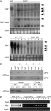
Effects of Temperature on Expression of Ugp1 and Ugp2 in Cosuppressing Plants. The numbers above the lanes indicate the following anther developmental stages: 1, premeiosis; 2, meiosis; 3, young microspore; 4, later microspore; and 5, pollen mitosis. Co27, cosuppressing plant of line 27; R19, RNAi plant of line 19. (A) RNA gel blot analysis of Ugp1 transcript levels in leaves of fertility-reverted cosuppressing plants at low temperature. The RNA gel blot was hybridized with the corresponding Ugp1 and subsequently with the Ubi1 intron probes. Arrowheads indicate (1) the unprocessed longer-than-full-length transcript and (2) endogenous Ugp1 mRNA; (3) to (5) indicate the silencing-related RNA degradation intermediates. Loading of equal amounts of RNA was confirmed by ethidium bromide staining. (B) RNA gel blot analysis of Ugp1 transcript levels in florets of fertility-reverted cosuppressing plants grown at low temperature. The RNA gel blot was hybridized with corresponding Ugp1 probes. Loading of equal amounts of RNA was confirmed by ethidium bromide staining. (C) Protein gel blot analysis of UGPase protein in cosuppressing plants. Protein samples were extracted from florets and leaves of cosuppressing plants grown at high temperature (male sterile stage) and low temperature (male fertile stage). (D) RT-PCR analysis of the expression of Ugp2 and correctly spliced Ugp1 from unprocessed primary transcripts derived from transcription of the Ugp1-OX construct (Figure 2A) in florets of cosuppressing plants. Amplification of the rice Actin1 gene was used as a control to show that approximately equal amounts of total RNA had been used in the RT-PCR analysis.

References

    1. Abe, T., Niiyama, H., and Sasahara, T. (2002). Cloning of cDNA for UDP-glucose pyrophosphorylase and the expression of mRNA in rice endosperm. Theor. Appl. Genet. 105 216–221. - PubMed
    1. Amor, Y., Haigler, C.H., Johnson, S., Wainscott, M., and Delmer, D.P. (1995). A membrane-associated form of sucrose synthase and its potential role in synthesis of cellulose and callose in plants. Proc. Natl. Acad. Sci. USA 92 9353–9357. - PMC - PubMed
    1. Arenas, J.E., and Abelson, J.N. (1997). Prp43: An RNA helicase-like factor involved in spliceosome disassembly. Proc. Natl. Acad. Sci. USA 94 11798–11802. - PMC - PubMed
    1. Baulcombe, D. (2004). RNA silencing in plants. Nature 431 356–363. - PubMed
    1. Beclin, C., Boutet, S., Waterhouse, P., and Vaucheret, H. (2002). A branched pathway for transgene-induced RNA silencing in plants. Curr. Biol. 12 684–688. - PubMed

Publication types

MeSH terms

Substances

Associated data

LinkOut - more resources