. 2007 Jul;56(7):1792-801.
doi: 10.2337/db06-1513. Epub 2007 Mar 30.

Glucose infusion in mice: a new model to induce beta-cell replication

Affiliations

Glucose infusion in mice: a new model to induce beta-cell replication

Laura C Alonso et al. Diabetes. 2007 Jul.

Abstract

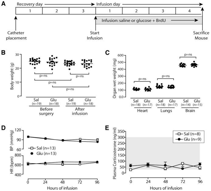
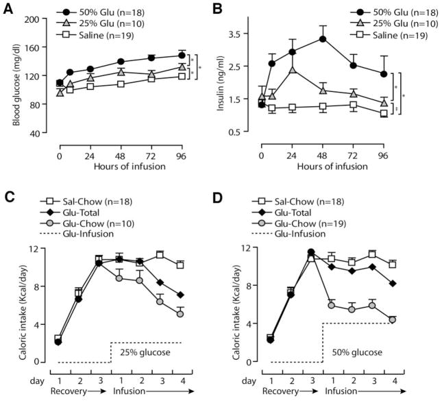
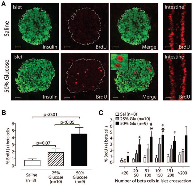
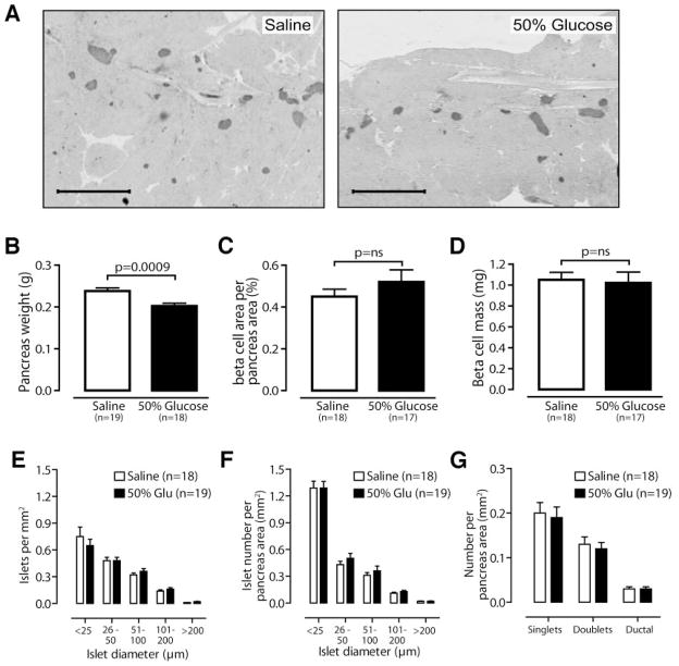
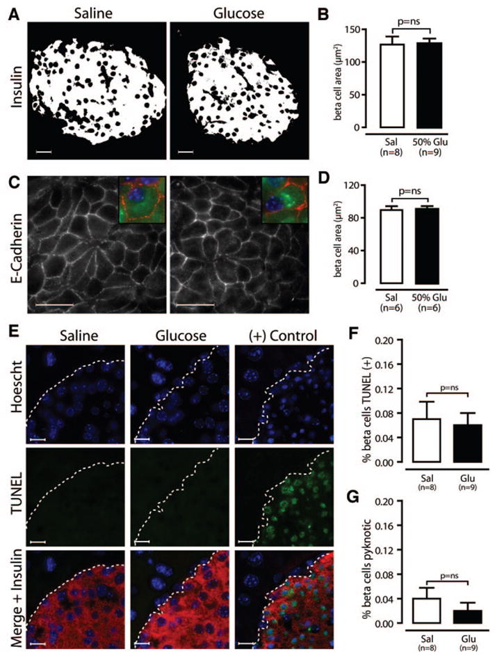
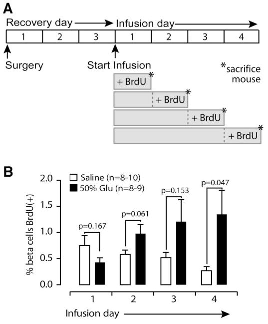
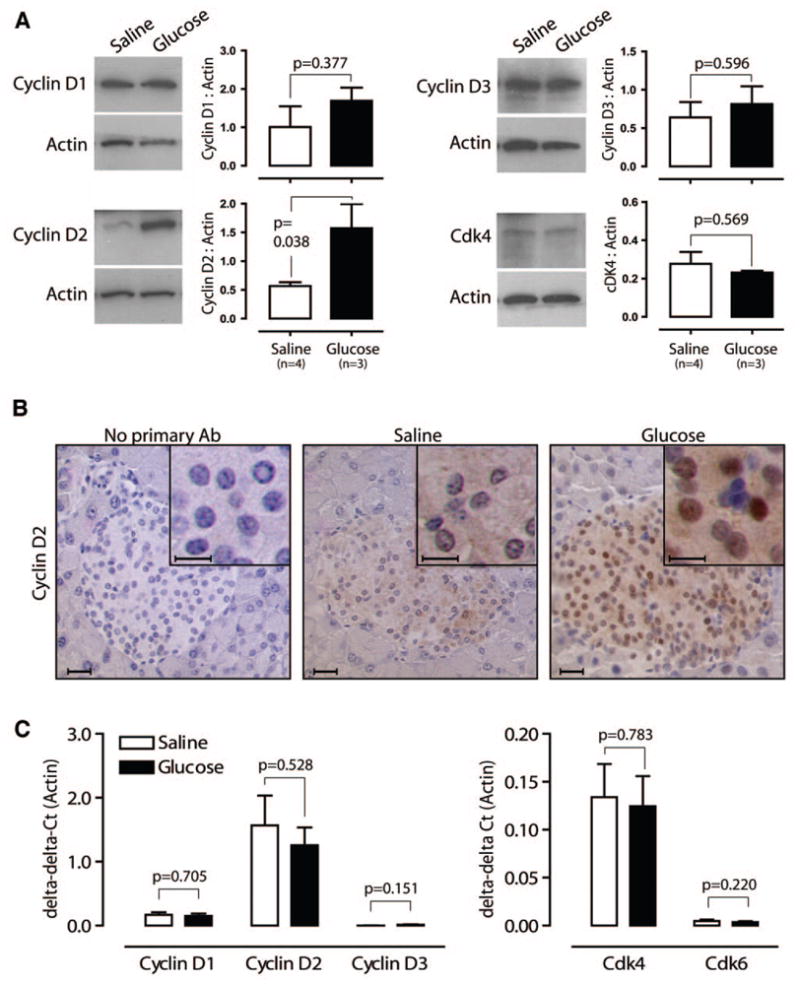
Developing new techniques to induce beta-cells to replicate is a major goal in diabetes research. Endogenous beta-cells replicate in response to metabolic changes, such as obesity and pregnancy, which increase insulin requirement. Mouse genetic models promise to reveal the pathways responsible for compensatory beta-cell replication. However, no simple, short-term, physiological replication stimulus exists to test mouse models for compensatory replication. Here, we present a new tool to induce beta-cell replication in living mice. Four-day glucose infusion is well tolerated by mice as measured by hemodynamics, body weight, organ weight, food intake, and corticosterone level. Mild sustained hyperglycemia and hyperinsulinemia induce a robust and significant fivefold increase in beta-cell replication. Glucose-induced beta-cell replication is dose and time dependent. Beta-cell mass, islet number, beta-cell size, and beta-cell death are not altered by glucose infusion over this time frame. Glucose infusion increases both the total protein abundance and nuclear localization of cyclin D2 in islets, which has not been previously reported. Thus, we have developed a new model to study the regulation of compensatory beta-cell replication, and we describe important novel characteristics of mouse beta-cell responses to glucose in the living pancreas.


Figures

FIG. 1
FIG. 1
Four-day glucose infusion is well tolerated in mice. A: Experiment timeline. Glucose infusion does not alter body weight (B), organ weight (C), or mean arterial blood pressure (BP) and heart rate (HR) (D). E: Plasma corticosterone is not elevated; gray area indicates normal range for mice (49). Data are means ± SE.
FIG. 2
FIG. 2
Glucose infusion induces mild metabolic alterations within the physiological range. Twenty-five and 50% glucose (Glu) significantly increase blood glucose (A) and plasma insulin (B). Sources of calories in 25% glucose-infused (C) and 50% glucose-infused (D) mice include infusate (dotted line) and chow (gray circles). Glucose-infused mice reduce chow intake, so total caloric intake (black diamonds) is similar to saline mice (white squares; saline data are repeated in C and D for comparison). Data are means ± SE; *P < 0.001, ‡P < 0.05 for one-way ANOVA using Tukey’s post-test comparison with saline.
FIG. 3
FIG. 3
Glucose infusion increases β-cell replication in a dose-dependent fashion. A: Representative islets stained for BrdU and insulin show glucose-induced replication; intestine is a positive control for BrdU exposure and staining. Inset: Confocal microscopy confirms BrdU-positive nuclei belong to insulin-positive cells. B: 50% glucose increases β-cell replication (vs. 25% glucose or saline); 25% glucose also shows a strong trend (vs. saline). C: Glucose (Glu) induces more replication in midsize islets than small islets. *P < 0.05 vs. saline; #P < 0.05 vs. “<20” islets. Scale bars = 20 μm; mean ± SE. Sal, saline.
FIG. 4
FIG. 4
β-Cell mass and islet number are unchanged after 4-day glucose infusion. A: Representative low-power images of pancreas sections stained for insulin and hematoxylin. B: Pancreas weight is reduced after glucose infusion. Percent β-cell area per pancreas area (C) and β-cell mass (D) are not increased in glucose-infused mice. Islet number is unchanged in glucose (Glu)-infused mice, whether counted digitally (E) or manually (FG) using a calibrated intraocular grid. G: The number of singlets, doublets, and insulin-positive ductal cells is unchanged in glucose-infused mice. Scale bars = 1 mm. Data are means ± SE.
FIG. 5
FIG. 5
β-Cell size and death are unchanged by glucose infusion. A and B: Integrated insulin area per β-cell is unchanged in glucose (Glu)-infused mice. C and D: Using sections stained for E-cadherin (red), insulin (green), and Hoechst (blue), measurement of β-cell area by tracing cell borders confirms that β-cell size is unchanged. E–G: Using sections stained for TUNEL (green), insulin (red), and Hoescht (blue), we find the rates of TUNEL (F) and pyknosis (G) are low in both groups. The positive control is a mouse treated with streptozotocin. Scale bars in A and C = 20 μm and in E = 10 μm. Data are means ± SE. Sal, saline.
FIG. 6
FIG. 6
Glucose-induced replication increases over 4-day infusion. A: Mice received 1, 2, 3, or 4 days of saline or 50% glucose, with BrdU added only for the final 24 h. B: Glucose (Glu)-induced replication increases during days 2, 3, and 4. Data are means ± SE.
FIG. 7
FIG. 7
Glucose infusion increases cyclin D2 abundance and nuclear localization. A: Islets from mice infused with saline or glucose for 4 days show that cyclin D2 protein abundance significantly increases; cyclin D1, cyclin D3, and cdk4 are unchanged. B: Immunohistochemistry for cyclin D2 reveals pronounced nuclear localization in islets from glucose-infused mice. C: Real-time PCR demonstrates no difference in mRNA abundance of D-type cyclins and cdks 4 and 6 in islets from saline- or glucose-infused mice. Scale bars = 20 μm (10 μm in insets). Data are means ± SE.

References

    1. Butler AE, Janson J, Bonner-Weir S, Ritzel R, Rizza RA, Butler PC. β-Cell deficit and increased β-cell apoptosis in humans with type 2 diabetes. Diabetes. 2003;52:102–110. - PubMed
    1. Yoon KH, Ko SH, Cho JH, Lee JM, Ahn YB, Song KH, Yoo SJ, Kang MI, Cha BY, Lee KW, Son HY, Kang SK, Kim HS, Lee IK, Bonner-Weir S. Selective beta-cell loss and alpha-cell expansion in patients with type 2 diabetes mellitus in Korea. J Clin Endocrinol Metab. 2003;88:2300–2308. - PubMed
    1. Bonner-Weir S, Weir GC. New sources of pancreatic beta-cells. Nat Biotechnol. 2005;23:857–861. - PubMed
    1. Bonner-Weir S, Deery D, Leahy JL, Weir GC. Compensatory growth of pancreatic β-cells in adult rats after short-term glucose infusion. Diabetes. 1989;38:49–53. - PubMed
    1. Lee HC, Bonner-Weir S, Weir GC, Leahy JL. Compensatory adaption to partial pancreatectomy in the rat. Endocrinology. 1989;124:1571–1575. - PubMed

Publication types