

doi: 10.1107/S1744309107006689.
Epub 2007 Mar 12.
Expression, crystallization and preliminary crystallographic data analysis of filamin A repeats 14-16
Affiliations
- PMID: 17401197
- PMCID: PMC2330200
- DOI: 10.1107/S1744309107006689
Item in Clipboard
Expression, crystallization and preliminary crystallographic data analysis of filamin A repeats 14-16
Acta Crystallogr Sect F Struct Biol Cryst Commun.
.
Abstract
Human filamin A is a 280 kDa protein involved in actin-filament cross-linking. It is structurally divided into an actin-binding headpiece (ABD) and a rod domain containing 24 immunoglobulin-like (Ig) repeats. A fragment of human filamin A (Ig repeats 14-16) was cloned and expressed in Escherichia coli and the purified protein was crystallized in 1.6 M ammonium sulfate, 2% PEG 1000 and 100 mM HEPES pH 7.5. The crystals diffracted to 1.95 A and belong to space group P2(1)2(1)2(1), with unit-cell parameters a = 50.63, b = 52.10, c = 98.46 A, alpha = beta = gamma = 90 degrees.
Figures
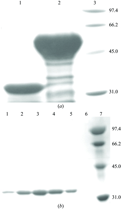
SDS–PAGE showing tag-cleavage and gel-filtration fractions of human filamin A repeats 14–16. (a) Cleaved protein (lane 1), uncleaved protein (lane 2) and molecular-weight standards (lane 3) with sizes labelled in kDa. (b) Fractions from gel filtration (lanes 1–6) showing the 36.3 kDa protein and molecular-weight standards (lane 7).
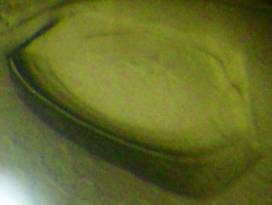
Crystal (dimensions 0.3 × 0.2 × 0.05 mm) of filamin A repeats 14–16 in 1.6 M ammonium sulfate, 2% PEG 1000 and 100 mM HEPES pH 7.5. Alhough lacking distinct edges, this lens-shaped crystal diffracted to 1.7 Å.
References
-
- Awata, H., Huang, C., Handlogten, M. E. & Miller, R. T. (2001). J. Biol. Chem.276, 34871–34879. - PubMed
-
- Carugo, K. D., Banuelos, S. & Saraste, M. (1997). Nature Struct. Biol.4, 175–179. - PubMed
-
- Collaborative Computational Project, Number 4 (1994). Acta Cryst. D50, 760–763. - PubMed
-
- Davies, P. J., Wallach, D., Willingham, M. C., Pastan, I., Yamaguchi, M. & Robson, R. M. (1978). J. Biol. Chem.253, 4036–4042. - PubMed
-
- Feng, Y. & Walsh, C. A. (2004). Nature Cell Biol.6, 1034–1038. - PubMed
Publication types
MeSH terms
Substances
LinkOut - more resources
Full Text Sources
