

Expression and purification of F-plasmid RepE and preliminary X-ray crystallographic study of its complex with operator DNA
- PMID: 17401213
- PMCID: PMC2330221
- DOI: 10.1107/S1744309107012894
Expression and purification of F-plasmid RepE and preliminary X-ray crystallographic study of its complex with operator DNA
Abstract
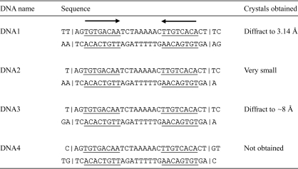
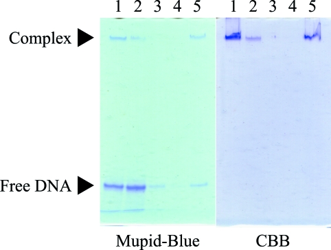
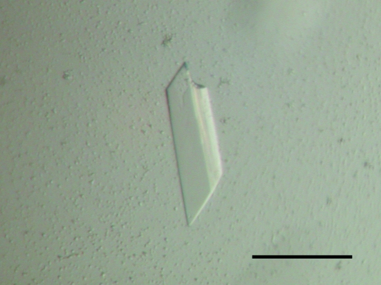
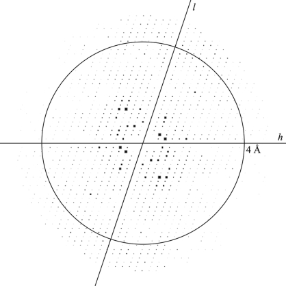
The replication initiator factor RepE of the F plasmid in Escherichia coli is an essential protein that stringently regulates the F-plasmid copy number. The RepE protein has a dual function: its monomer functions as a replication initiator, while its dimer acts as a transcriptional repressor of the repE gene. The wild-type dimeric RepE protein was expressed as an N-terminal histidine-tagged protein, purified under native conditions with a high salt concentration and crystallized in complex with the repE operator DNA using the sitting-drop vapour-diffusion technique. The crystals diffracted to a resolution of 3.14 A after the application of dehydration and crystal annealing and belong to space group P2(1), with unit-cell parameters a = 60.73, b = 99.32, c = 95.00 A, beta = 108.55 degrees.
Figures
Similar articles
-
Regulation of DNA replication by iterons: an interaction between the ori2 and incC regions mediated by RepE-bound iterons inhibits DNA replication of mini-F plasmid in Escherichia coli.EMBO J. 1999 Jul 1;18(13):3856-67. doi: 10.1093/emboj/18.13.3856. EMBO J. 1999. PMID: 10393200 Free PMC article.
-
Crystal structure of a prokaryotic replication initiator protein bound to DNA at 2.6 A resolution.EMBO J. 1999 Sep 1;18(17):4597-607. doi: 10.1093/emboj/18.17.4597. EMBO J. 1999. PMID: 10469640 Free PMC article.
-
The N-terminal domain of the replication initiator protein RepE is a dimerization domain forming a stable dimer.Biochem Biophys Res Commun. 2004 Feb 27;315(1):10-5. doi: 10.1016/j.bbrc.2004.01.018. Biochem Biophys Res Commun. 2004. PMID: 15013418
-
Replication initiator protein RepE of mini-F plasmid: functional differentiation between monomers (initiator) and dimers (autogenous repressor).Proc Natl Acad Sci U S A. 1994 Apr 26;91(9):3839-43. doi: 10.1073/pnas.91.9.3839. Proc Natl Acad Sci U S A. 1994. PMID: 8170998 Free PMC article.
-
Twenty years of the pPS10 replicon: insights on the molecular mechanism for the activation of DNA replication in iteron-containing bacterial plasmids.Plasmid. 2004 Sep;52(2):69-83. doi: 10.1016/j.plasmid.2004.06.002. Plasmid. 2004. PMID: 15336485 Review.
Cited by
-
Measurement of the equilibrium relative humidity for common precipitant concentrations: facilitating controlled dehydration experiments.Acta Crystallogr Sect F Struct Biol Cryst Commun. 2012 Jan 1;68(Pt 1):111-4. doi: 10.1107/S1744309111054029. Epub 2011 Dec 24. Acta Crystallogr Sect F Struct Biol Cryst Commun. 2012. PMID: 22232186 Free PMC article.
-
Increasing the X-ray diffraction power of protein crystals by dehydration: the case of bovine serum albumin and a survey of literature data.Int J Mol Sci. 2012;13(3):3782-3800. doi: 10.3390/ijms13033782. Epub 2012 Mar 21. Int J Mol Sci. 2012. PMID: 22489183 Free PMC article.
-
Replisome Assembly at Bacterial Chromosomes and Iteron Plasmids.Front Mol Biosci. 2016 Aug 11;3:39. doi: 10.3389/fmolb.2016.00039. eCollection 2016. Front Mol Biosci. 2016. PMID: 27563644 Free PMC article. Review.
-
Structural basis for regulation of bifunctional roles in replication initiator protein.Proc Natl Acad Sci U S A. 2007 Nov 20;104(47):18484-9. doi: 10.1073/pnas.0705623104. Epub 2007 Nov 13. Proc Natl Acad Sci U S A. 2007. PMID: 18000058 Free PMC article.
-
Raoult's law revisited: accurately predicting equilibrium relative humidity points for humidity control experiments.J Appl Crystallogr. 2017 Mar 29;50(Pt 2):631-638. doi: 10.1107/S1600576717003636. eCollection 2017 Apr 1. J Appl Crystallogr. 2017. PMID: 28381983 Free PMC article.
References
-
- Collaborative Computational Project, Number 4 (1994). Acta Cryst. D50, 760–763. - PubMed
-
- Harp, J. M., Timm, D. E. & Bunick, G. J. (1998). Acta Cryst. D54, 622–628. - PubMed
-
- Heras, B. & Martin, J. L. (2005). Acta Cryst. D61, 1173–1180. - PubMed
-
- Joachimiak, A. & Sigler, P. B. (1991). Methods Enzymol.208, 82–99. - PubMed
Publication types
MeSH terms
Substances
LinkOut - more resources
Full Text Sources
Other Literature Sources
