Review
. 2007 Apr;8(4):335-9.
doi: 10.1038/sj.embor.7400942.

The emergence of shape: notions from the study of the Drosophila tracheal system

Affiliations
Review

The emergence of shape: notions from the study of the Drosophila tracheal system

Jordi Casanova. EMBO Rep. 2007 Apr.

Abstract

The generation of bodies and body parts with specific shapes and sizes has been a longstanding issue in biology. Morphogenesis in general and organogenesis in particular are complex events that involve global changes in cell populations in terms of their proliferation, migration, differentiation and shape. Recent studies have begun to address how these synchronized changes are controlled by the genes that specify cell fate and by the ability of cells to respond to extracellular cues. In particular, a notable shift in this research has occurred owing to the ability to address these issues in the context of the whole organism. For such studies, the Drosophila tracheal system has proven to be a particularly appropriate model. Here, my aim is to highlight some ideas that have arisen through our studies, and those from other groups, of Drosophila tracheal development. Rather than providing an objective review of the features of tracheal development, I intend to discuss some selected notions that I think are relevant to the question of shape generation.


Figures
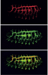
Figure 1
Figure 1
The tracheal system of the Drosophila embryo. The embryonic Drosophila tracheal system as visualized with the 2A12 antibody (top), which recognizes a lumen component, and with a GFP antibody (middle) in an embryo carrying a UAS–tau-GFP reporter construct under the control of the tracheal-specific btl–GAL4 driver. A merged image is also shown (bottom). The tracheal system consists of different branches on the antero-posterior and dorso-ventral axes, each with a stereotypical pattern and size. Anterior is on the left and dorsal is at the top. Images provided by S. Araujo. GFP, green fluorescent protein.
Figure 2
Figure 2
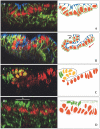
Interfaces and forces that shape body parts. (A,B) The tracheal cells (nuclei are shown in red) are selected to invaginate, whereas the neighbouring ectodermal cells remain at the embryonic surface. Tracheal cells are labelled using a Trh antibody. Anti-neurotactin labels the basolateral and basal sides of all epithelial cells (shown in green), whereas protein kinase C (PKC) labels their apical side (shown in blue). The arrows in (B) indicate the direction of movement. (C) Among the invaginating tracheal cells, the dorsal cells that express the gene sal begin a rotation-like movement, whereas the ventral cells slide below the dorsal ones. The tracheal cells are labelled as described in (A) and are shown in red, whereas the sal-positive cells are detected with a specific antibody (shown in green); tracheal sal-positive cells are visualized in yellow. (D) Accumulation of myosin II is detected at the onset of invagination in the cells initiating apical constriction (shown by the white line and asterisk), as visualized by a green fluorescent protein-tagged form of the myosin II light chain. Tracheal cells are labelled as described in (A) and are shown in red. Figures modified with permission from Brodu & Casanova, 2006.
Figure 3
Figure 3
Constraints, cast and size. (A,B) The tracheal cells of the dorsal branches (DB; shown in black) are located between the precursors of the dorsal oblique (DO) muscles (shown in brown). Note that by stage 14, one can observe several tracheal cells in the same row (A), whereas by stage 15, on intercalation, they form a one-cell row (B). (C) The tracheal cells of the lateral trunk anterior and posterior (Lta and Ltp, respectively; shown in black) form two branches in apposition to the lateral transverse (LT) and ventral acute (VA) muscles (shown in brown). Note that the shapes of Lta and Ltp result from their close apposition to the mesodermal cells. (D) The shape of the visceral branch results from the apposition of the tracheal cells (shown in red) to the cells of the visceral mesoderm (vm; shown in green). (E) In the dorsal trunk (DT; shown in the antero-posterior axis) two or more cells contribute to the tube circumference. Conversely, in the DB (shown in the dorso-ventral axis), the tube circumference is made from single cells wrapped around the lumen. In (AC), mesoderm cells are visualized in embryos carrying the twi-CD2 gene stained with an antibody against CD2, and the tracheal cells are visualized with the antibody against Trunk (Trk) in embryos carrying tracheal-specific btl–GAL4 and UAS–trk constructs. Images modified with permission from Franch-Marro & Casanova (2000). In (D), tracheal cells are visualized with a β-galactosidase antibody in embryos from the 1-eve-1 enhancer trap line, and the visceral mesoderm is detected by a FasIII antibody. Image modified with permission from Boube et al (2001). In (E), tracheal cells are detected by tracheal expression of a tau-GFP construct and the tracheal lumen by wheat germ agglutinin (Araujo et al, 2005). GFP, green fluorescent protein.
None
Jordi Casanova

References

    1. Affolter M, Shilo B (2000) Genetic control of branching morphogenesis during Drosophila tracheal development. Curr Opin Cell Biol 12: 731–735 - PubMed
    1. Affolter M, Bellusci S, Itoh N, Shilo B, Thiery J-P, Werb Z (2003) Tube or not tube: remodeling epithelial tissues by branching morphogenesis. Dev Cell 4: 11–18 - PubMed
    1. Araujo SJ, Aslam H, Tear G, Casanova J (2005) mummy/cystic encodes for an enzyme required for chitin and glycan synthesis, involved in trachea, embryonic cuticle and CNS development: analysis of its role in Drosophila tracheal morphogenesis. Dev Biol 288: 179–193 - PubMed
    1. Baumgartner S, Littleton JT, Broadie K, Bhat MA, Harbecke R, Lengyel JA, Chiquet-Ehrismann R, Prokop A, Bellen HJ (1996) A Drosophila neurexin is required for septate junction and blood–nerve barrier formation and function. Cell 87: 1059–1068 - PubMed
    1. Behr M, Riedel D, Schuh R (2003) The claudin-like megatrachea is essential in septate junctions for the epithelial barrier function in Drosophila. Dev Cell 5: 611–620 - PubMed

Publication types

LinkOut - more resources