. 2007 Jun;101(5):1205-13.
doi: 10.1111/j.1471-4159.2007.04487.x. Epub 2007 Mar 30.

Glutamate release from activated microglia requires the oxidative burst and lipid peroxidation

Affiliations

Glutamate release from activated microglia requires the oxidative burst and lipid peroxidation

Steven W Barger et al. J Neurochem. 2007 Jun.

Abstract

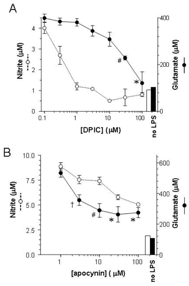
When activated by proinflammatory stimuli, microglia release substantial levels of glutamate, and mounting evidence suggests this contributes to neuronal damage during neuroinflammation. Prior studies indicated a role for the Xc exchange system, an amino acid transporter that antiports glutamate for cystine. Because cystine is used for synthesis of glutathione (GSH) synthesis, we hypothesized that glutamate release is an indirect consequence of GSH depletion by the respiratory burst, which produces superoxide from NADPH oxidase. Microglial glutamate release triggered by lipopolysaccharide was blocked by diphenylene iodonium chloride and apocynin, inhibitors of NADPH oxidase. This glutamate release was also blocked by vitamin E and elicited by lipid peroxidation products 4-hydroxynonenal and acrolein, suggesting that lipid peroxidation makes crucial demands on GSH. Although NADPH oxidase inhibitors also suppressed nitrite accumulation, vitamin E did not; moreover, glutamate release was largely unaffected by nitric oxide donors, inhibitors of nitric oxide synthase, or changes in gene expression. These findings indicate that a considerable degree of the neurodegenerative consequences of neuroinflammation may result from conversion of oxidative stress to excitotoxic stress. This phenomenon entails a biochemical chain of events initiated by a programmed oxidative stress and resultant mass-action amino acid transport. Indeed, some of the neuroprotective effects of antioxidants may be due to interference with these events rather than direct protection against neuronal oxidation.


Figures

Figure 1
Figure 1. Antioxidant effects on microglial glutamate release
Primary microglia were treated with vitamin E or NAC for 30 min prior to application of LPS (100 ng/ml). After 20 h, medium was harvested for assay of nitrite (A only) and glutamate. In A, vitamin E was 100 μM and NAC was 500 μM. Values represent the means ± SEM of quadruplicate (A) or triplicate (B) cultures. Effects of vitamin E on glutamate were significant by ANOVA and Scheffe post hoc test (*p<0.01 vs. LPS alone).
Figure 2
Figure 2. Lipid peroxidation products evoke glutamate release
Primary microglia were treated with acrolein or 4-HNE alone at the indicated concentrations; after 20 h glutamate levels were measured. Values represent mean ± SEM of quadruplicate cultures. All responses above 3 μM were significant (*p<0.0001, #p<0.002 vs. control) by ANOVA and Scheffe post hoc test.
Figure 3
Figure 3. Effect of NADPH oxidase inhibition in microglial glutamate release
Primary microglia were treated with DPIC (A) or apocynin (B) for 30 min prior to application of LPS (100 ng/ml). After 20 h, medium was harvested for assay of nitrite and glutamate. Values represent the means ± SEM of quadruplicate (A) or triplicate (B) cultures. Bars represent levels of nitrite (open) and glutamate (filled) in untreated cultures. Effects of DPIC and apocynin on glutamate were significant by ANOVA and Scheffe post hoc test (*p<0.0002, #p<0.02, p<0.05 vs. LPS alone).
Figure 4
Figure 4. Tests of a role for nitric oxide in microglial glutamate release
Primary microglia were treated with NO donors (A) or LPS (30 ng/ml) with and without inhibitors of NO synthase (B). After 20 h, medium was harvested for assay of glutamate. Values represent the means ± SEM of quadruplicate cultures. The glutamate levels in untreated cultures (open bar, B) or in cultures treated with LPS alone (black bars) are also indicated. Abbr.: NaNP, sodium nitroprusside; SNAP, S-Nitroso-N-acetylpenicillamine; SIN-1, 3-morpholinosydnonimine; nitro-Arg, Nω-nitro-L-arginine; N-propyl-Arg, N-propyl-L-arginine. No significant effects of the NO donors or NOS inhibitors was found by ANOVA and Scheffe post hoc test.
Figure 5
Figure 5. Glutamate release is independent of macromolecular synthesis
A: Cycloheximide or α-amanitin was applied to primary microglia for 1 h prior to the application of LPS (30 ng/ml). After 16 h, medium was collected for assay of glutamate. The glutamate levels in untreated cultures (white bar) or in cultures treated with LPS alone (black bar) are also indicated. Values represent mean ± SEM of quadruplicate cultures. The effect of α-amanitin was significant by ANOVA and Scheffe post hoc (p<0.02). B: The indicated inhibitors were applied to primary microglia in triplicate for 1 h prior to the application of 30 ng/ml LPS; “JNK inh” = SP600125 (30 μM), “MEK inh” = U0126 (10 μM), α-AA = α-amino adipate (2.5 mM). After 20 h, medium was collected for assay of glutamate, and the cells were harvested for preparation of RNA. Solid bars represent glutamate values as mean ± SEM (# p<0.05 vs. LPS alone). Stipled bars indicate the level of xCT mRNA relative to 18S rRNA in each sample; values represent mean ± SEM (* p<0.02 vs. LPS alone).
Figure 6
Figure 6. Hypothetical chain of events connecting NADPH oxidase to glutamate release
Evidence from other studies indicates that PKC activation and phosphorylation of p47phox are important events in the activation of NADPH oxidase by proinflammatory stimuli. Once assembled at the membrane, p47phox and the other components of NADPH oxidase produce superoxide. This is dismutated by SOD to hydrogen peroxide. Some of the peroxide is reduced by glutathione peroxidase, consuming GSH. Some peroxide putatively initiates lipid oxidation, which must be halted by covalent conjugation of GSH to the lipid by glutathione S-transferase, particularly GSTA4-4. The depletion of GSH resulting from these events is replaced by de novo synthesis, requiring cysteine, imported into the cell as cystine via the Xc exchange mechanism. Though not required for the initiation of this mechanism, events mediated by JNK and ERK can also elevate expression of the antiporter’s xCT subunit. Pharmacological agents shown in the present work to block these events are enclosed in red boxes; entry into the chain of events at the point of lipid peroxidation is highlighted by the green box.

References

    1. Alblas J, Honing H, de Lavalette CR, Brown MH, Dijkstra CD, van den Berg TK. Signal regulatory protein alpha ligation induces macrophage nitric oxide production through JAK/STAT- and phosphatidylinositol 3-kinase/Rac1/NAPDH oxidase/H2O2-dependent pathways. Mol Cell Biol. 2005;25:7181–7192. - PMC - PubMed
    1. Awasthi YC, Yang Y, Tiwari NK, Patrick B, Sharma A, Li J, Awasthi S. Regulation of 4-hydroxynonenal-mediated signaling by glutathione S-transferases. Free Radic Biol Med. 2004;37:607–619. - PubMed
    1. Bal-Price A, Moneer Z, Brown GC. Nitric oxide induces rapid, calcium-dependent release of vesicular glutamate and ATP from cultured rat astrocytes. Glia. 2002;40:312–323. - PubMed
    1. Barger SW. An unconventional hypothesis of oxidation in Alzheimer’s disease: intersections with excitotoxicity. Front Biosci. 2004;9:3286–3295. - PubMed
    1. Barger SW, Basile AS. Activation of microglia by secreted amyloid precursor protein evokes release of glutamate by cystine exchange and attenuates synaptic function. J Neurochem. 2001;76:846–854. - PubMed

Publication types