. 2007 Apr 15;21(8):904-19.
doi: 10.1101/gad.1516607. Epub 2007 Apr 2.

Discovery of tMAC: a Drosophila testis-specific meiotic arrest complex paralogous to Myb-Muv B

Affiliations

Discovery of tMAC: a Drosophila testis-specific meiotic arrest complex paralogous to Myb-Muv B

Eileen L Beall et al. Genes Dev. .

Abstract

The Drosophila Myb-Muv B (MMB)/dREAM complex regulates gene expression and DNA replication site-specifically, but its activities in vivo have not been thoroughly explored. In ovarian amplification-stage follicle cell nuclei, the largest subunit, Mip130, is a negative regulator of replication, whereas another subunit, Myb, is a positive regulator. Here, we identified a mutation in mip40 and generated a mutation in mip120, two additional MMB subunits. Both mutants were viable, but mip120 mutants had many complex phenotypes including shortened longevity and severe eye defects. mip40 mutant females had severely reduced fertility, whereas mip120 mutant females were sterile, substantiating ovarian regulatory role(s) for MMB. Myb accumulation and binding to polytene chromosomes was dependent on the core factors of the MMB complex. In contrast to the documented mip130 mutant phenotypes, both mip40 and mip120 mutant males were sterile. We purified Mip40-containing complexes from testis nuclear extracts and identified tMAC, a new testis-specific meiotic arrest complex that contained Mip40, Caf1/p55, the Mip130 family member, Always early (Aly), and a Mip120 family member, Tombola (Tomb). Together, these data demonstrate that MMB serves diverse roles in different developmental pathways, and members of MMB can be found in alternative, noninteracting complexes in different cell types.


Figures

Figure 1.
Figure 1.
Molecular verification of the mip40 and mip120 mutants. (A) The chromosomal configuration of the mip40 locus is shown (purple and pink), along with the location of the P-element insert in the mip40-coding sequence (orange double-sided arrow). (B) The chromosomal configuration of the mip120 locus is shown, with the mip120 exons in pink. The location of eftu-M (CG6050) within the first mip120 intron is shown in green. The P-element insertion for line KG05422 is shown in orange. The location of the probe used in C (blue) and the EcoRI restriction sites are also shown. The two lines generated after mobilization of the P-element and used in this study, eftu-M34 and mip12067, are shown. The regions deleted in these mutants are indicated by the red arrows. (C) Southern blot of eftu-M34 and mip12067 containing a transgene expressing eftu-M on the third chromosome (P[eftu-M]). Shown are EcoRI digests of DNA isolated from the strains indicated on the top and probed with the DNA fragment shown in B. The size of the deletion in each mutant as determined by sequence analysis is as follows: eftu-M34, 2236 bp; mip12067, 5770 bp. (D) Whole animals of the indicated genotypes were homogenized in sample buffer. “e2f2” refers to flies derived from crossing e2f2329/CyoKrGFP and e2f278A/CyoKrGFP (Cayirlioglu et al. 2001). Fly equivalents (0.3) were loaded onto a 9% SDS-PAGE, immunoblotted, and analyzed for the accumulation of the indicated MMB subunit using affinity-purified antibodies. Tubulin served as the loading control.
Figure 2.
Figure 2.
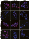
Chromatin association of Myb is dependent on Mip120 and Mip130. Confocal images of polytene chromosome spreads using GFP-sorted wandering third instar larvae from the following strains are shown: mip12067-9A-9/CyOKrGFP, mip1301–723 + 1–36/FM7KrGFP or mybMH107/FM7cAcGFP. The control (GFP+) or mutant (GFP−) spreads are as indicated on the left. (Blue) DAPI; (red) Anti-Mip120, Anti-Mip130, or Anti-Myb as indicated on the top.
Figure 3.
Figure 3.
Mip40 and Mip120 are required for egg chamber development. (A,B,E,F) Shown are merged confocal images of stage 10 egg chambers from females of the genotypes indicated on the bottom of each panel. (Red) Anti-Orc2; (green) Anti-BrdU; (blue) DAPI. (C,D) Same as in other panels, except that a lower-magnification view (10×) is shown to demonstrate the lack of stage 10 and later egg chambers in mip120 mutants.
Figure 4.
Figure 4.
The MMB complex is present at ACE3 prior to, and during, chorion gene amplification. Sheared cross-linked chromatin from preamplification (less than or equal to stage 9) and amplification (stage 10) egg chambers was used for ChIP using the indicated antibodies. Shown is an ethidium bromide-stained gel of PCR reactions performed using both ACE3-specific and actin-specific (control) primers simultaneously in the reaction containing either input or immunoprecipitated DNA. ACE3 is enriched for all samples except the control IgG and input samples. Note that the ratio of ACE3:actin is higher in the input DNA for amplification-stage egg chambers (lane 8) when compared with preamplification egg chambers (lane 1) due to higher copy numbers of the ACE3 locus in this sample.
Figure 5.
Figure 5.
Mip40 and Mip120 are required for male fertility. Shown are phase-contrast images using a Leica DM5000 microscope of testes dissected from mip40 (A–C) or mip120 (D–F) males of the genotypes indicated in each panel. (B) Note the complete lack of mature sperm in mip40EY16520 mutants due to presumed arrest at the spermatocyte stage of spermatid differentiation. Arrows identify sperm bundles present in control (D) and mip120 mutant (E) testes.
Figure 6.
Figure 6.
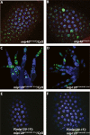
Mip40 and Mip120 proteins are expressed in spermatocytes. Shown are merged confocal images of testes from wild-type males stained with the antibodies indicated on the top (red). (Green) Anti-BrdU; (blue) DAPI. (A–C) View (10×) of whole testis to show staining in the mitotic (green) and spermatocyte stages. (D–E) Higher-magnification image (25×) to show detail.
Figure 7.
Figure 7.
Meiotic arrest proteins specifically interact with Mip40. (A) Poros-heparin fractions of Drosophila testis extracts were immunoprecipitated using the antibodies indicated at the top. The precipitates were washed with buffer containing 0.5 M KCl, and the coimmunoprecipitated proteins were eluted with 0.4% sarcosyl. As a result of the sarcosyl wash, the protein to which the antibody was raised will remain in the pellet and will not appear in the IP supernatant. All immunoprecipitations were performed in the presence of 50 μg/mL ethidium bromide. Shown are the immunoblot analyses of dissected whole testes, ovaries, and the immunoprecipitated material using the antibodies indicated on the left. Only Mip40 antibodies coimmunoprecipitated Aly and Comr specifically. It also appears that MMB is an abundant complex relative to tMAC, based on the qualitative signal strengths of immunoblot signals for Aly and Comr versus Mip40 and Myb in the testis lane. (B) Data from duplicate control and Mip40 antibody columns were analyzed by mass spectroscopy. Above the dotted line is a list of MMB subunits identified by mass spectrometry from the Mip40 antibody column eluate that were not present in the control antibody column eluate. In addition, several testis-specific proteins that have known functions in meiotic arrest were also identified only in the Mip40 eluate, including Aly, Comr, Tomb, and Topi. (C) Drosophila Tomb belongs to the Tesmin/TSO family of proteins. Shown is an alignment of the CXC domain of Tomb and other tesmin-family proteins: Human Tesmin (AAH64579), Dog Tesmin (XP_854573), Mouse Tesmin (BAB64935), Drosophila Mip120, Drosophila Tombola, and Arabidopsis SOL1. (*) Conserved residues; (:) similar residues among the aligned proteins.
Figure 8.
Figure 8.
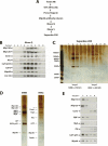
Mip40 and Aly are present in a newly identified complex in testes. (A) The fractionation scheme used to purify tMAC. Fractions from each chromatography step were immunoblotted for Mip40, Comr, and Aly. Peak fractions were pooled and used for the subsequent chromatography step. (B) Aly and Comr polypeptides cofractionated with MMB using ion-exchange chromatography. Shown are immunoblots of the Mono Q fractions indicated on the top of the panel using antibodies directed against Aly, Comr, and subunits of the MMB complex as indicated on the left. (C) Fractions eluting from the final Superdex 200 gel filtration column were resolved on a 4%–12% gradient SDS-PAGE and visualized by silver staining. The peaks of MMB (fractions 2,3) and tMAC (fractions 7,8) are indicated at the bottom. The MMB and tMA complexes eluted with an estimated molecular weight of ∼700 kDa and ∼400 kDa, respectively. Molecular weight markers are indicated on the left. (D) The peaks of MMB (fraction 2) and tMAC (fraction 7) from C are compared. Proteins identified by size and confirmed by immunoblot analysis (see below) are indicated on the left of each panel. Bands labeled as Topi and Tomb are based on the predicted molecular weight of the full-length polypeptides. The band marked with a “?” represents an unknown cofractionating protein that is not Mip120. (E) The only MMB proteins present in tMAC are Mip40 and Caf1/p55. Shown are immunoblots of the Superdex 200 gel filtration fractions indicated at the top using antibodies against the MMB complex subunits Aly and Comr as indicated on the left.

Similar articles

Cited by

References

    1. Ashburner M. Drosophila: A laboratory manual. Cold Spring Harbor Laboratory Press; Cold Spring Harbor, NY: 1989.
    1. Beall E.L., Manak J.R., Zhou S., Bell M., Lipsick J.S., Botchan M.R., Manak J.R., Zhou S., Bell M., Lipsick J.S., Botchan M.R., Zhou S., Bell M., Lipsick J.S., Botchan M.R., Bell M., Lipsick J.S., Botchan M.R., Lipsick J.S., Botchan M.R., Botchan M.R. Role for a Drosophila Myb-containing protein complex in site-specific DNA replication. Nature. 2002;420:833–837. - PubMed
    1. Beall E.L., Bell M., Georlette D., Botchan M.R., Bell M., Georlette D., Botchan M.R., Georlette D., Botchan M.R., Botchan M.R. Dm-myb mutant lethality in Drosophila is dependent upon mip130: Positive and negative regulation of DNA replication. Genes & Dev. 2004;18:1667–1680. - PMC - PubMed
    1. Boxem M., van den Heuvel S., van den Heuvel S. C. elegans class B synthetic multivulva genes act in G(1) regulation. Curr. Biol. 2002;12:906–911. - PubMed
    1. Calvi B.R., Lilly M.A., Spradling A.C., Lilly M.A., Spradling A.C., Spradling A.C. Cell cycle control of chorion gene amplification. Genes & Dev. 1998;12:734–744. - PMC - PubMed

Publication types

MeSH terms

Substances

LinkOut - more resources