. 2007 Apr 1;21(7):744-9.
doi: 10.1101/gad.1519107.

The microRNA miR-124 antagonizes the anti-neural REST/SCP1 pathway during embryonic CNS development

Affiliations

The microRNA miR-124 antagonizes the anti-neural REST/SCP1 pathway during embryonic CNS development

Jaya Visvanathan et al. Genes Dev. .

Abstract

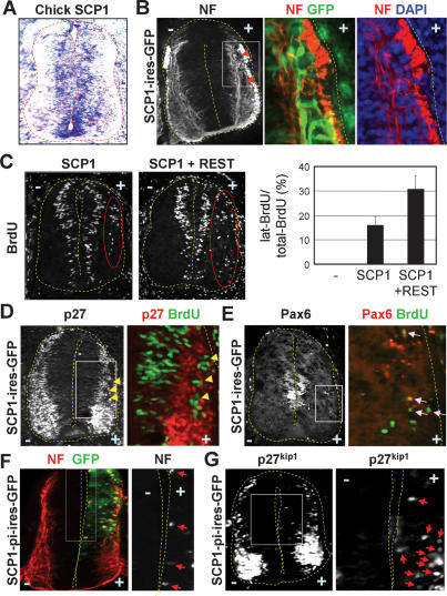
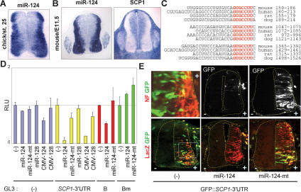
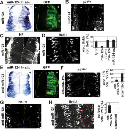
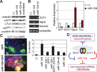
Neuronal gene expression is tightly regulated in developing CNS. Here, we demonstrate the anti-neural function of phosphatase SCP1 (small C-terminal domain phosphatase 1) during development. We further show that the neuron-enriched microRNA miR-124 directly targets SCP1-3' untranslated region (UTR) to suppress SCP1 expression. In developing spinal cord, expression of miR-124 and SCP1 is complementary, and miR-124 antagonism phenocopies SCP1 overexpression and vice versa. In P19 cells, miR-124 suppresses SCP1 expression and induces neurogenesis, and SCP1 counteracts this proneural activity of miR-124. Our results suggest that, during CNS development, timely down-regulation of SCP1 is critical for inducing neurogenesis, and miR-124 contributes to this process at least in part by down-regulating SCP1 expression.


Figures

Figure 1.
Figure 1.
SCP1 as an anti-neural factor in the chick neural tube. (A) In situ hybridization on chick embryos reveals that SCP1 is widely expressed but down-regulated in the lateral neuronal zone. (B) SCP1-misexpressing cells (GFP+ cells) in the lateral zone lack the NF expression. (C) Misexpression of SCP1 leads to ectopic BrdU+-proliferating cells in the lateral zone (cells in red circle), and this is markedly enhanced by coexpression of REST. (D,E) While ectopic BrdU+ cells lack post-mitotic marker p27kip1 (arrows in D), they maintain expression of progenitor gene Pax6 (arrows in E). (F,G) Phosphatase-inactive SCP1 mutant SCP1-pi induces ectopic and precocious NF+ neuronal differentiation (arrows in F) and expression of p27kip1 (arrows in G) in the medial zone of the chick neural tube.
Figure 2.
Figure 2.
SCP1-3′UTR as a direct target of miR-124. (A,B) In situ detection of miR-124 in chick and mouse embryos shows that miR-124 is highly and specifically expressed in the lateral neuronal zone of the neural tube and DRG, complementary to the expression pattern of SCP1. (C) Three evolutionarily conserved miR-124 target sites are found at the 3′UTR of SCP1 mRNA. Seed match sequences are marked in red. (D) Luciferase assays with the GL3∷SCP1-3′UTR reporter in HEK293 cells. Both synthetic miR-124 RNA duplexes (miR-124) and miR-124 expression vector (CMV-124) repress luciferase expression from GL3∷SCP1-3′UTR, while the miR-124 mutant and miR-128 have no effect. GL3 vector alone is minimally affected by miRNAs. The B-segment of SCP1-3′UTR containing the second miR-124 target site is suppressed by miR-124, and mutation on the miR-124 target site in the B-segment (Bm) abolishes the repression by miR-124. (E) Electroporation of GFP∷SCP1-3′UTR directs a high level of GFP expression in the medial zone and a lower GFP expression in the NF+ lateral zone of the chick neural tube. Coelectroporation of miR-124 diminishes GFP expression from GFP∷SCP1-3′UTR, whereas miR-124-mt has no effect. CMV-LacZ was included as an electroporation indicator.
Figure 3.
Figure 3.
Proneural activity of miR-124 in the chick neural tube. (A–D) Overexpression of miR-124 in the chick spinal cord. (A) Electroporation of miR-124 RNA duplexes (with CMV-GFP as an electroporation indicator) increases miR-124 expression in the developing chick spinal cord 2 d post-electroporation as detected with in situ hybridization. (B–D) miR-124 overexpression triggers ectopic neurogenesis (p27kip1+, NF+ post-mitotic neuronal cells in B,C) in the more medial area of the neural tube, accompanied by compromised progenitor cell proliferation (D). (E–H) Inhibition of miR-124 function in the chick spinal cord. Electroporation of 2′-OMe antisense miR124 (anti-miR-124) with CMV-GFP interferes with miR-124 detection by in situ hybridization (E), decreases the expression of neuronal markers p27kip1 and NeuN in the lateral zone of the neural tube (F,G), and simultaneously triggers laterally positioned BrdU+-proliferating cells (cells in red circle in H).
Figure 4.
Figure 4.
miR-124-induced neurogenesis is antagonized by SCP1 in P19 mouse embryonic cells. (A) Western analyses show that, in P19 cells, miR-124 expression induces post-mitotic cell marker p27kip1 and neuronal marker TuJ, while suppressing proliferation marker cyclinA. Notably, transfection indicator GFP expression is lower in miR-124-transfected cells. (B) RT–PCR assays reveal that expression of miR-124, but not miR-124-mt, reduces SCP1 and induces proneural bHLH factors Ngn2 and NeuroD and neuronal marker TuJ. Quantitative RT–PCR results are as shown. (C) Neuronal differentiation analyses in transfected P19 cells. miR-124 expression triggers TuJ+ neuronal differentiation, which is compromised by cotransfection of the miR-124-resistant SCP1 vector but not by the miR-124-sensitive SCP1 vector containing SCP1-3′UTR (SCP1∷ 3′UTR). Percentage of neuronal differentiation is measured by percentage of TuJ+ neurons among GFP+-transfected cells. (D) Negative feedback loop between the anti-neural REST/SCP1 and proneural miR-124 pathways contributes to a rapid and efficient transition of cellular phenotypes between neural progenitors and post-mitotic neurons (see text). Blue and red denote the regulatory pathways in nonneurons and neurons, respectively.

Similar articles

Cited by

References

    1. Alvarez-Garcia I., Miska E.A., Miska E.A. MicroRNA functions in animal development and human disease. Development. 2005;132:4653–4662. - PubMed
    1. Ambros V. The functions of animal microRNAs. Nature. 2004;431:350–355. - PubMed
    1. Andres M.E., Burger C., Peral-Rubio M.J., Battaglioli E., Anderson M.E., Grimes J., Dallman J., Ballas N., Mandel G., Burger C., Peral-Rubio M.J., Battaglioli E., Anderson M.E., Grimes J., Dallman J., Ballas N., Mandel G., Peral-Rubio M.J., Battaglioli E., Anderson M.E., Grimes J., Dallman J., Ballas N., Mandel G., Battaglioli E., Anderson M.E., Grimes J., Dallman J., Ballas N., Mandel G., Anderson M.E., Grimes J., Dallman J., Ballas N., Mandel G., Grimes J., Dallman J., Ballas N., Mandel G., Dallman J., Ballas N., Mandel G., Ballas N., Mandel G., Mandel G. CoREST: A functional corepressor required for regulation of neural-specific gene expression. Proc. Natl. Acad. Sci. 1999;96:9873–9878. - PMC - PubMed
    1. Ballas N., Battaglioli E., Atouf F., Andres M.E., Chenoweth J., Anderson M.E., Burger C., Moniwa M., Davie J.R., Bowers W.J., Battaglioli E., Atouf F., Andres M.E., Chenoweth J., Anderson M.E., Burger C., Moniwa M., Davie J.R., Bowers W.J., Atouf F., Andres M.E., Chenoweth J., Anderson M.E., Burger C., Moniwa M., Davie J.R., Bowers W.J., Andres M.E., Chenoweth J., Anderson M.E., Burger C., Moniwa M., Davie J.R., Bowers W.J., Chenoweth J., Anderson M.E., Burger C., Moniwa M., Davie J.R., Bowers W.J., Anderson M.E., Burger C., Moniwa M., Davie J.R., Bowers W.J., Burger C., Moniwa M., Davie J.R., Bowers W.J., Moniwa M., Davie J.R., Bowers W.J., Davie J.R., Bowers W.J., Bowers W.J., et al. Regulation of neuronal traits by a novel transcriptional complex. Neuron. 2001;31:353–365. - PubMed
    1. Ballas N., Grunseich C., Lu D.D., Speh J.C., Mandel G., Grunseich C., Lu D.D., Speh J.C., Mandel G., Lu D.D., Speh J.C., Mandel G., Speh J.C., Mandel G., Mandel G. REST and its corepressors mediate plasticity of neuronal gene chromatin throughout neurogenesis. Cell. 2005;121:645–657. - PubMed

Publication types

MeSH terms