. 2007 Apr 1;21(7):756-69.
doi: 10.1101/gad.1535507.

Expansion of adult beta-cell mass in response to increased metabolic demand is dependent on HNF-4alpha

Affiliations

Expansion of adult beta-cell mass in response to increased metabolic demand is dependent on HNF-4alpha

Rana K Gupta et al. Genes Dev. .

Abstract

The failure to expand functional pancreatic beta-cell mass in response to increased metabolic demand is a hallmark of type 2 diabetes. Lineage tracing studies indicate that replication of existing beta-cells is the principle mechanism for beta-cell expansion in adult mice. Here we demonstrate that the proliferative response of beta-cells is dependent on the orphan nuclear receptor hepatocyte nuclear factor-4alpha (HNF-4alpha), the gene that is mutated in Maturity-Onset Diabetes of the Young 1 (MODY1). Computational analysis of microarray expression profiles from isolated islets of mice lacking HNF-4alpha in pancreatic beta-cells reveals that HNF-4alpha regulates selected genes in the beta-cell, many of which are involved in proliferation. Using a physiological model of beta-cell expansion, we show that HNF-4alpha is required for beta-cell replication and the activation of the Ras/ERK signaling cascade in islets. This phenotype correlates with the down-regulation of suppression of tumorigenicity 5 (ST5) in HNF-4alpha mutants, which we identify as a novel regulator of ERK phosphorylation in beta-cells and a direct transcriptional target of HNF-4alpha in vivo. Together, these results indicate that HNF-4alpha is essential for the physiological expansion of adult beta-cell mass in response to increased metabolic demand.


Figures
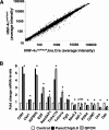
Figure 1.
Figure 1.
Gene expression profiling of HNF-4αloxP/loxP;Ins.Cre mice. Total RNA was prepared from isolated islets of 4- to 5-mo-old control and mutant mice. The RNA was amplified, fluorescently labeled, and hybridized to the PancChip 5.0 cDNA microarray. (A) A scatterplot of average intensity values for all expressed genes illustrates that the vast majority of genes were not differentially expressed. Approximately 2% of expressed genes (128 genes significantly up-regulated and 57 significantly down-regulated) were differentially expressed in HNF-4α mutant islets (n = 5 HNF-4αloxP/loxP, n = 3 HNF-4αloxP/loxP;Ins.Cre). (B) Quantitative real-time PCR confirmation of differentially expressed genes. Total RNA was extracted from isolated islets of a second cohort of animals, and cDNA derived from unamplified RNA was used for PCR. Thirteen of 14 (92%) genes were confirmed as differentially expressed, consistent with the 10% FDR setting chosen for microarray analysis (n = 4–6 for PCR). (*) p-value from Student’s t-test <0.05.
Figure 2.
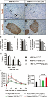
Figure 2.
HNF-4α is required for β-cell expansion during pregnancy. BrdU staining of pancreata from 4- to 5-mo-old pregnant HNF-4αloxP/loxP (A) and HNF-4αloxP/loxP;Ins.Cre (B) mice. (C,D) Insulin staining of adjacent sections indicates β-cells within the islet. (E) Percentage of islet nuclei staining positive for BrdU is lower in pregnant HNF-4αloxP/loxP;Ins.Cre (white) mice when compared with pregnant HNF-4αloxP/loxP (black) and HNF-4α+/+;Ins.Cre (gray) controls. (F) β-cell mass of pregnant HNF-4αloxP/loxP;Ins.Cre (white) mice is significantly lower than that of pregnant HNF-4αloxP/loxP (black) and HNF-4α+/+;Ins.Cre (gray) controls. (G) Consistent with morphometric analysis, total pancreatic insulin content in pregnant HNF-4αloxP/loxP;Ins. Cre (white) mice is reduced when compared with pregnant HNF-4αloxP/loxP (black) and HNF-4α+/+;Ins.Cre (gray) controls. (H) Distribution of islet size in HNF-4αloxP/loxP (black) and HNF-4αloxP/loxP;Ins.Cre (white) mice indicates fewer larger-sized islets in pregnant HNF-4α mutants. Bars represent the mean ± SEM; n = 3–4 mice for each group for AH. (*) p-value from Student’s t-test <0.05. (I) Glucose tolerance tests of virgin and pregnant HNF-4αloxP/loxP and HNF-4αloxP/loxP; Ins.Cre mice. The area below the curve measurements (0–30 min) indicates that the impact of HNF-4α deficiency on glucose tolerance is more pronounced in the pregnant state. The data represent the mean ± SEM; n = 6–8 mice for each group. (*) p-value from Student’s t-test <0.05 when compared with pregnant controls.
Figure 3.
Figure 3.
Ras/ERK signaling in β-cells is HNF-4α dependent. (A) Ras-GTP ELISA indicates lower levels of activated Ras in freshly isolated islets from virgin HNF-4α mutant mice in comparison to controls; n = 3 for each group. (B) Western blot analysis indicates that EGF stimulation of ERK phosphorylation in isolated islets is HNF-4α dependent. VAMP2 (vesicle-associated membrane protein 2) expression was used as a loading control. (C) Western blot analysis indicates that in freshly isolated islets of control mice, ERK1 and ERK2 phosphorylation increases dramatically during pregnancy but is significantly lower in pregnant HNF-4α-deficient islets. (D) Real-time PCR analysis confirms microarray results indicating reduced mRNA levels of the ERK activator ST5 in isolated islets of HNF-4α mutants; n = 6 mice for each group. (E) Adenoviral transduction of MIN6 cells with shRNA targeting ST5 results in 70% reduction in ST5 mRNA levels; n = 4 for each group. (F) Western blot analysis indicates that MIN6 cells with reduced expression of ST5 exhibit lower levels of basal and EGF-stimulated phosphorylated ERK. VAMP2 expression was used as a loading control. (*) p-value from Student’s t-test <0.05.
Figure 4.
Figure 4.
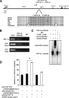
ST5 is a direct transcriptional target of HNF-4α in vivo. (A) Genomic organization of the mouse ST5 locus with the evolutionarily conserved HNF-4α-binding site in intron 1. The position of the binding site is indicated relative to the transcriptional start site (TSS). Arrows indicate the location of primer pairs used for Chip assays. (B) ChIP assays from isolated adult islets using an antibody raised against HNF-4α reveal that HNF-4α occupies the intronic region of ST5 containing the high-scoring HNF-4α-binding site (conserved in humans) but does not occupy the putative site −55 kb upstream of the promoter. Primers amplifying the known HNF-4α-binding site in the HNF-1α promoter serve as the positive control for the HNF-4α ChIP assay. (C) EMSA. Incubation of liver nuclear extract with a radiolabeled oligonucleotide containing the putative HNF-4α-binding sequence at intron 1 resulted in a strong shift of the radioactive band. The intensity of this band was diminished with the addition of an unlabeled competitor oligonucleotide containing a known HNF-4α-binding site. Addition of a HNF-4α antibody generated a supershifted band that was not observed with the addition of preimmune serum, confirming the identity of the bound protein as HNF-4α. (D) Cotransfection of BHK cells with HNF-4α and pGL3-ST5, expressing luciferase under the control of a 192-bp fragment of the 3′ conserved region of ST5 containing the HNF-4α-binding site, results in increased luciferase activity, indicating that this element serves as an HNF-4α-dependent enhancer. Mutation of this binding site nearly abolishes the transcriptional activation. (*) p-value < 0.05 by ANOVA; n = 3 for each transfection condition.
Figure 5.
Figure 5.
Combining location and expression microarray analysis in pancreatic islets. (A) Venn diagram displaying the overlap of orthologous genes between the two platforms compared in this analysis. The genes on the PancChip cDNA array are represented as the white circle, while the genes on the human promoter array are shown in black. Of the cDNAs represented on the PancChip, 4755 (the gray intersect) have orthologs for which the proximal promoter is present on the human promoter chip developed by Odom et al. (2004). (B) Venn diagram displaying the overlap of RNA polymerase II-bound promoters (black circles) and genes expressed in pancreatic islets identified by expression profiling (white circles). (C) Venn diagram displaying the overlap of HNF4α-bound promoters (black circle) in pancreatic islets and genes differentially expressed (white) in islets lacking HNF4α in β-cells. (D) Histogram of the absolute change in gene expression (either repression or induction) for 505 genes identified in the location analysis by Odom et al. (2004).

References

    1. Ahlgren U., Jonsson J., Jonsson L., Simu K., Edlund H., Jonsson J., Jonsson L., Simu K., Edlund H., Jonsson L., Simu K., Edlund H., Simu K., Edlund H., Edlund H. β-Cell-specific inactivation of the mouse Ipf1/Pdx1 gene results in loss of the β-cell phenotype and maturity onset diabetes. Genes & Dev. 1998;12:1763–1768. - PMC - PubMed
    1. Amaral M.E., Ueno M., Carvalheira J.B., Carneiro E.M., Velloso L.A., Saad M.J., Boschero A.C., Ueno M., Carvalheira J.B., Carneiro E.M., Velloso L.A., Saad M.J., Boschero A.C., Carvalheira J.B., Carneiro E.M., Velloso L.A., Saad M.J., Boschero A.C., Carneiro E.M., Velloso L.A., Saad M.J., Boschero A.C., Velloso L.A., Saad M.J., Boschero A.C., Saad M.J., Boschero A.C., Boschero A.C. Prolactin-signal transduction in neonatal rat pancreatic islets and interaction with the insulin-signaling pathway. Horm. Metab. Res. 2003;35:282–289. - PubMed
    1. Amaral M.E., Cunha D.A., Anhe G.F., Ueno M., Carneiro E.M., Velloso L.A., Bordin S., Boschero A.C., Cunha D.A., Anhe G.F., Ueno M., Carneiro E.M., Velloso L.A., Bordin S., Boschero A.C., Anhe G.F., Ueno M., Carneiro E.M., Velloso L.A., Bordin S., Boschero A.C., Ueno M., Carneiro E.M., Velloso L.A., Bordin S., Boschero A.C., Carneiro E.M., Velloso L.A., Bordin S., Boschero A.C., Velloso L.A., Bordin S., Boschero A.C., Bordin S., Boschero A.C., Boschero A.C. Participation of prolactin receptors and phosphatidylinositol 3-kinase and MAP kinase pathways in the increase in pancreatic islet mass and sensitivity to glucose during pregnancy. J. Endocrinol. 2004;183:469–476. - PubMed
    1. Blake J.A., Richardson J.E., Bult C.J., Kadin J.A., Eppig J.T., Richardson J.E., Bult C.J., Kadin J.A., Eppig J.T., Bult C.J., Kadin J.A., Eppig J.T., Kadin J.A., Eppig J.T., Eppig J.T. MGD: The Mouse Genome Database. Nucleic Acids Res. 2003;31:193–195. - PMC - PubMed
    1. Briancon N., Weiss M.C., Weiss M.C. In vivo role of the HNF4α AF-1 activation domain revealed by exon swapping. EMBO J. 2006;25:1253–1262. - PMC - PubMed

Publication types

MeSH terms

LinkOut - more resources