. 2007 Apr 1;21(7):821-34.
doi: 10.1101/gad.1536107.

U2 toggles iteratively between the stem IIa and stem IIc conformations to promote pre-mRNA splicing

Affiliations

U2 toggles iteratively between the stem IIa and stem IIc conformations to promote pre-mRNA splicing

Angela K Hilliker et al. Genes Dev. .

Abstract

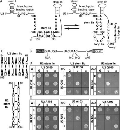
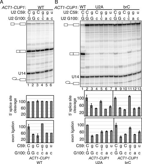
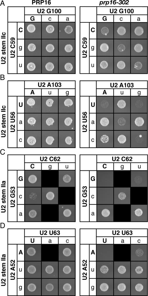
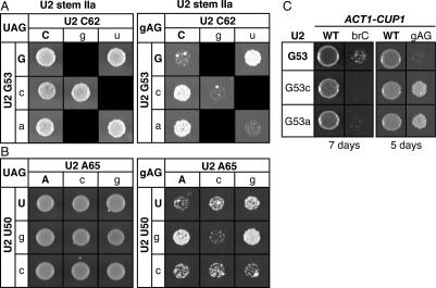
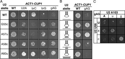
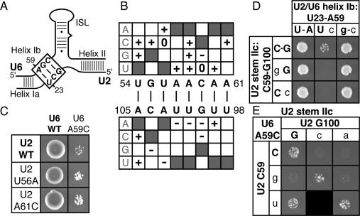
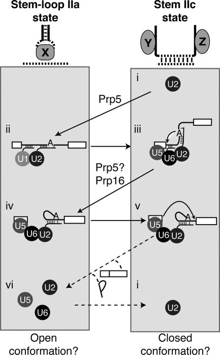
To ligate exons in pre-messenger RNA (pre-mRNA) splicing, the spliceosome must reposition the substrate after cleaving the 5' splice site. Because spliceosomal small nuclear RNAs (snRNAs) bind the substrate, snRNA structures may rearrange to reposition the substrate. However, such rearrangements have remained undefined. Although U2 stem IIc inhibits binding of U2 snRNP to pre-mRNA during assembly, we found that weakening U2 stem IIc suppressed a mutation in prp16, a DExD/H box ATPase that promotes splicing after 5' splice site cleavage. The prp16 mutation was also suppressed by mutations flanking stem IIc, suggesting that Prp16p facilitates a switch from stem IIc to the mutually exclusive U2 stem IIa, which activates binding of U2 to pre-mRNA during assembly. Providing evidence that stem IIa switches back to stem IIc before exon ligation, disrupting stem IIa suppressed 3' splice site mutations, and disrupting stem IIc impaired exon ligation. Disrupting stem IIc also exacerbated the 5' splice site cleavage defects of certain substrate mutations, suggesting a parallel role for stem IIc at both catalytic stages. We propose that U2, much like the ribosome, toggles between two conformations--a closed stem IIc conformation that promotes catalysis and an open stem IIa conformation that promotes substrate binding and release.


Figures

Figure 1.
Figure 1.
Disrupting U2 stem IIc exacerbates the splicing defect of a substrate mutated at the branch site or the 5′ splice site. (A) Structure of U2 stem IIc and stem–loop IIa. Nucleotides involved in stem IIc, stem IIa, and loop IIa are bold. The nucleotides of U2 that bind the intronic branch site consensus sequence are shown. Nucleotide sequence and numbers correspond to the Saccharomyces cerevisiae sequence. The dashed line marks a nonconserved base pair that can extend stem IIa in S. cerevisiae. (B) A pictogram of stem IIc and IIa showing the consensus sequence for organisms ranging from budding yeast to humans; numbering refers to the budding yeast sequence. The height of the letter is proportional to the frequency of the nucleotide in the alignment. (C) Schematic diagram of the ACT1-CUP1 splicing reporter, which when spliced confers copper resistance to budding yeast (Lesser and Guthrie 1993). The intron consensus sequences for budding yeast are shown along with point mutations used in this study. The branch site adenosine is bold. (D,E) Mutations that disrupt base-pairing in U2 stem IIc enhance the growth defect conferred by the 5′ splice site mutation U2A and the branch site mutation brC. Compensatory analysis by copper resistance of U2 stem IIc base pairs C59/G100 (D) and U56/A103 (E) is shown. The matrices show the copper resistance of the wild-type (WT; left), brC (middle), or U2A (right) ACT1-CUP1 splicing reporters in yJPS1035 expressing U2 variants having single or double mutations in the stem IIc base pairs. Cells were grown for 3–4 d at 30°C on solid media containing 0.05 mM copper sulfate. Wild-type residues are capitalized.
Figure 2.
Figure 2.
Disrupting U2 stem IIc exacerbates the 5′ splice site cleavage defect of a substrate mutated at the branch site or the 5′ splice site and impairs exon ligation of a wild-type substrate. (A,B) Compensatory analysis by splicing in vivo of the U2 stem IIc base pair C59/G100 is shown. Primer extension analysis shows the in vivo splicing phenotype of wild-type (WT, A) or mutated U2A and brC (B) ACT1-CUP1 splicing reporters in yJPS1035 having single or double mutations in the stem IIc base pair. The pre-mRNA, mRNA, and lariat intermediate of the ACT1-CUP1 reporter and U14, which serves as an internal control, are highlighted. Quantitation of the primer extensions is shown below. The identities of the bases are indicated above the gel and below the graph; wild-type residues are capitalized. The apparent efficiency of 5′ splice site cleavage is calculated as (mRNA + lariat intermediate)/(pre-mRNA + lariat intermediate + mRNA). The apparent efficiency of exon ligation is calculated as (mRNA/lariat intermediate). Values are normalized to the strain expressing wild-type U2. The histograms show the mean of duplicate samples; the error bars indicate the range of values. While the trends of the exon ligation efficiencies in A repeated in independent experiments, the magnitude of the differences varied.
Figure 3.
Figure 3.
Disrupting U2 stem IIc or flanking structures mutually exclusive with stem IIa suppresses a mutation in PRP16, a DExD/H box ATPase. (A,B) Disruption of U2 stem IIc suppresses the mutation prp16-302. Compensatory analysis by growth of the U2 stem IIc base pairs C59/G100 (A) and U56/A103 (B) in a wild-type PRP16 (left) or a mutant prp16-302 (right) strain is shown. The cells were grown for 3 d (left) or 6 d (right) at 20°C on solid media containing 5-FOA. (C,D) Disruption of structures mutually exclusive with stem IIa suppresses prp16-302. Compensatory analysis by growth of the U2 stem IIa base pairs G53/C62 (C) and A52/U63 (D) in a wild-type PRP16 (left) or a mutant prp16-302 (right) strain. Cells were grown for 3 d (D, left), 4 d (C, left) or 6 d (C,D, right) at 20°C on solid media containing 5-FOA. Black boxes indicate that the mutation is lethal and could not be tested. Matrices are labeled as in Figure 1.
Figure 4.
Figure 4.
Destabilization of stem IIa suppresses the 3′ splice site mutant gAG. (A,B) Disruption of base-pairing in U2 stem IIa suppresses a 3′ splice site mutation. Compensatory analysis by copper resistance of the U2 stem IIa base pair G53/C62 (A) or U2–U50/A65 (B) is shown. The matrices show the copper resistance of a wild-type (left) or mutated 3′ splice site mutant (gAG; right) ACT1-CUP1 splicing reporter in yJPS1035 expressing a U2 variant having single or double mutations in the stem IIa base pairs. Cells were grown for 3 d (left) or 5 d (right) at 30°C on solid media containing 0.05 mM CuSO4. Black boxes represent lethal U2 alleles that could not be included in the analysis. (C) Disrupting U2 stem IIa does not suppress the mutated branch site substrate brC. For comparison, suppression of the gAG mutated 3′ splice site substrate is shown. Mutations were tested in strain yJPS1035. The identity of U2 is indicated to the left and the identity of the reporter is indicated at the top. Cells were grown for 5 or 7 d, as indicated below the data, at 30°C on 0.1 mM CuSO4.
Figure 5.
Figure 5.
Mutations in U2 loop IIa suppress the 3′ splice site mutation gAG and the branch site mutation brG, independently of disrupting stem IIc. (AC) Copper resistance of U2 mutants in yJPS1035 expressing the wild-type, U2A, brC, brG, or gAG ACT1-CUP1 reporters. Cells were grown at 30°C on solid media containing 0.2 mM (A,C) or 0.1 mM (B) CuSO4. (A) Point mutants in loop IIa suppress the 3′ splice site mutation gAG and the branch site mutation brG but exacerbate or fail to suppress the U2A and brC mutations. Cells were grown for 2 d (WT; U2A), 3 d (gAG), 4 d (brC), or 5 d (brG). For each strain, the identity of U2 is indicated to the left (cf. Fig. 1A) and the identity of the ACT1-CUP1 reporter is shown at the top. (B) Mutations that severely alter loop IIa suppress gAG strongly, but mutations that severely alter the downstream strand of stem IIc do not. The position of the mutations in loop IIa or the downstream strand of stem IIc is indicated by asterisks below the affected strand. The U2 alleles are (from top row to bottom) wild type, UAA56–58AUU, GUAA57–58CAUU, UUA101–103AAU, and UUGUUACA98–105AACAAUGU. Cells were grown for 3 d (WT) or 6 d (gAG) at 30°C on solid media containing 0.2 mM CuSO4. (C) Restoration of stem IIc does not abolish suppression but rather improves suppression. A compensatory analysis by copper resistance of the U2 stem IIc base pair U56/A103 is shown. The matrix shows the copper resistance of the gAG mutated 3′ splice site reporter in yJP1035 expressing U2 variants having single or double mutations in the stem IIc base pair. Cells were grown for 6 d at 0.1 mM CuSO4.
Figure 6.
Figure 6.
Disrupting U2 stem IIc exacerbates disruption of U2/U6 helix Ib, which includes the U6–AGC triad. (A) Schematic of U2/U6 base-pairing; U2/U6 helix Ib is boxed. (BE) Mutations in the 5′ side of stem IIc largely suppress U6–A59C, while mutations in the 3′ side largely exacerbate. Genetic interactions between U2 stem IIc point or double mutations and U6 variants in strain XYU96 (Xu et al. 1998) are shown. (B) Summary of genetic interactions between U2 stem IIc point mutations and U6–A59C, a mutation in the AGC triad that disrupts U2/U6 helix Ib. Weak suppressors (+), enhancers (−), and those that do not interact (0) are indicated. Shaded boxes mark wild-type nucleotides; white boxes mark mutations that were not tested. Note: For all mutations tested, repair of stem IIc failed to abolish suppression (data not shown) but did abolish enhancement (see E; data not shown). (C) Representative mutations in loop IIa that weakly suppress the growth defect of U6–A59C. The identity of U2 is indicated to the right and the identity of U6 is indicated on top. Cells were grown for 6 d at 30°C on solid media containing 5-FOA. (D) Mutations in U2 stem IIc that exacerbate the growth defect of U6–A59C do so by enhancing disruption of U2/U6 helix Ib. The matrix shows the growth of cells expressing U2 variants that disrupt the U2 stem IIc base pair C59/G100 (rows) and U2 and/or U6 mutations that alter base-pairing in U2/U6 helix Ib (columns). Cells were grown for 5 d at 30°C on solid media containing 5-FOA. (E) Mutations in U2 stem IIc that exacerbate the growth defect of U6–A59C do so by disrupting stem IIc. A compensatory analysis by growth is shown for the U2 stem IIc base pair C59/G100. All strains expressed U6–A59C. Growth conditions were the same as in D.
Figure 7.
Figure 7.
U2 stem–loop IIa and stem IIc toggle during both the assembly phase and the catalytic phase of splicing. Our data suggest a model in which U2 stem II toggles between stem–loop IIa and stem IIc throughout splicing; this toggling may reflect larger conformational rearrangements in the spliceosome. We propose that the stem IIc state stabilizes a closed state of the spliceosome that promotes catalysis (right conformation states) and that stem–loop IIa stabilizes an open state of the spliceosome that promotes substrate sampling, rearrangement, and release (left conformational states). First, the free U2 snRNP in the stem IIc state (i) is converted by the DExD/H box ATPase Prp5p to the stem–loop IIa state to promote binding of U2 to the pre-mRNA and formation of the prespliceosome (Perriman and Ares 2007). Next, U2 must toggle from the stem–loop IIa state back to the stem IIc state, as the stem IIc state promotes 5′ splice site cleavage (iii; Figs. 1, 2). After 5′ splice site cleavage, stem IIc is destabilized by the DExD/H box ATPase Prp16p (Fig. 3), directly or indirectly, promoting formation of stem–loop IIa in the inferred intermediate (iv), a reaction that may also be promoted by Prp5p (Perriman and Ares 2007). Stem–loop IIa, in turn, is destabilized (Figs. 4, 5) to promote reformation of stem IIc and exon ligation (v; Figs. 2, 5C, 6). After exon ligation, toggling of stem IIc back to stem–loop IIa (vi) could reconfigure the spliceosome to promote product release and/or spliceosome disassembly. The free U2 snRNP may then switch back to the stem IIc state (i). The connectivity or changing connectivity of the substrate is shown for each conformation, and the snRNPs are shown as circles. Our data suggest that loop IIa interacts with an unknown factor, shown here as “X,” in the intermediate (iv; Figs. 5, 6) and that the strands of stem IIa interact with unknown factors, shown here as “Y” and “Z,” in the 5′ splice site cleavage conformation (ii; Fig. 3C,D), precluding stem IIa formation at this stage.

References

    1. Ansari A., Schwer B., Schwer B. SLU7 and a novel activity, SSF1, act during the PRP16-dependent step of yeast pre-mRNA splicing. EMBO J. 1995;16:4001–4009. - PMC - PubMed
    1. Ares M., Jr., Igel A.H., Igel A.H. Lethal and temperature-sensitive mutations and their suppressors identify an essential structural element in U2 small nuclear RNA. Genes & Dev. 1990;12A:2132–2145. - PubMed
    1. Barabino S.M., Sproat B.S., Lamond A.I., Sproat B.S., Lamond A.I., Lamond A.I. Antisense probes targeted to an internal domain in U2 snRNP specifically inhibit the second step of pre-mRNA splicing. Nucleic Acids Res. 1992;17:4457–4464. - PMC - PubMed
    1. Brow D.A., Guthrie C., Guthrie C. Spliceosomal RNA U6 is remarkably conserved from yeast to mammals. Nature. 1988;6179:213–218. - PubMed
    1. Brys A., Schwer B., Schwer B. Requirement for SLU7 in yeast pre-mRNA splicing is dictated by the distance between the branchpoint and the 3′ splice site. RNA. 1996;7:707–717. - PMC - PubMed

Publication types