. 2007 Jun;27(11):4018-27.
doi: 10.1128/MCB.01839-06. Epub 2007 Apr 2.

Eos, MITF, and PU.1 recruit corepressors to osteoclast-specific genes in committed myeloid progenitors

Affiliations

Eos, MITF, and PU.1 recruit corepressors to osteoclast-specific genes in committed myeloid progenitors

Rong Hu et al. Mol Cell Biol. 2007 Jun.

Abstract

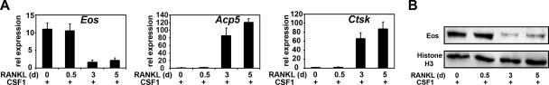
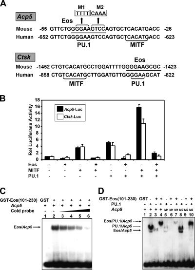
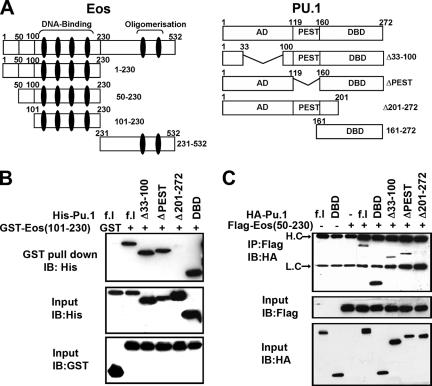
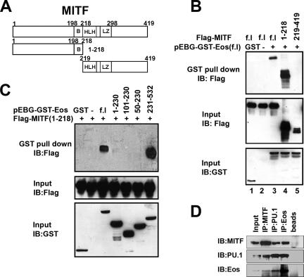
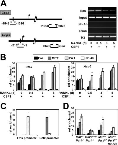
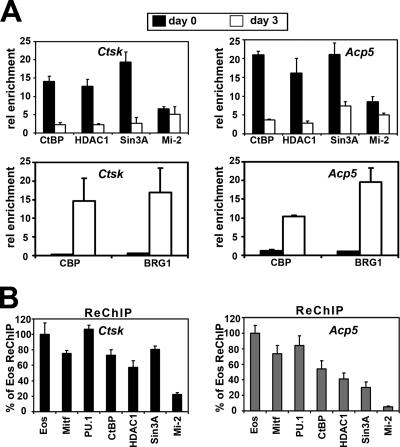
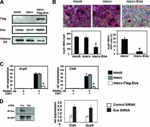
Transcription factors MITF and PU.1 collaborate to increase expression of target genes like cathepsin K (Ctsk) and acid phosphatase 5 (Acp5) during osteoclast differentiation. We show that these factors can also repress transcription of target genes in committed myeloid precursors capable of forming either macrophages or osteoclasts. The direct interaction of MITF and PU.1 with the zinc finger protein Eos, an Ikaros family member, was necessary for repression of Ctsk and Acp5. Eos formed a complex with MITF and PU.1 at target gene promoters and suppressed transcription through recruitment of corepressors CtBP (C-terminal binding protein) and Sin3A, but during osteoclast differentiation, Eos association with Ctsk and Acp5 promoters was significantly decreased. Subsequently, MITF and PU.1 recruited coactivators to these target genes, resulting in robust expression of target genes. Overexpression of Eos in bone marrow-derived precursors disrupted osteoclast differentiation and selectively repressed transcription of MITF/PU.1 targets, while small interfering RNA knockdown of Eos resulted in increased basal expression of Ctsk and Acp5. This work provides a mechanism to account for the modulation of MITF and PU.1 activity in committed myeloid progenitors prior to the initiation of osteoclast differentiation in response to the appropriate extracellular signals.


Figures

FIG. 1.
FIG. 1.
Eos expression is downregulated during osteoclast differentiation. (A) Relative (rel) expression of Eos, Acp5, and Ctsk mRNA was measured by qRT-PCR at the indicated times (in days [d]) and cytokine treatments. Results from three independent experiments are presented as means plus standard errors of the means (error bars). (B) Nuclear extracts from osteoclasts harvested at the indicated times (in days [d]) were analyzed by Western blotting using anti-Eos antibody. Histone H3 was used as a loading control.
FIG. 2.
FIG. 2.
Eos represses both Acp5 and Ctsk promoter activity. (A) Acp5 and Ctsk promoter sequences from mouse and human cells with conserved MITF and PU.1 binding sites (shown by brackets), and the putative Eos binding site (underlined). M1 and M2 show the sequence replacements within the Eos DNA consensus, as indicated. (B) Transient-transfection assays in NIH 3T3 cells. Either Acp5 luciferase reporter (Acp5-luc) or the Ctsk luciferase reporter (Ctsk-luc) was transfected alone or together with indicated combinations of expression vectors encoding MITF, PU.1, and Eos. Total DNA in each transfection was kept constant by adding empty expression vector. Relative (Rel) luciferase activity was represented as the difference from basal promoter activity (n-fold) (set at 1). Results from three independent experiments are presented as means plus standard errors of the means (error bars). (C) EMSAs using γ-32P end-labeled Acp5 oligonucleotide and recombinant GST or GST-Eos (101-230) protein. The formation of the DNA-Eos complex (arrow) was competed with increasing amounts of cold Acp5 probe. (D) EMSAs using γ-32P end-labeled wild-type or mutated Acp5 oligonucleotides in the presence of recombinant His6-PU.1 and GST-Eos (101-230) protein. The Eos-DNA complex (thick arrow), PU.1-DNA complex (thin arrow), and the supershifted band containing both Eos and PU.1 (broken arrow) are indicated.
FIG. 3.
FIG. 3.
Physical interaction of Eos with PU.1. (A) Schematic representation of Eos and PU.1 domains and the respective deletion mutations used in the present study. Zinc fingers in Eos are represented as a black vertical eclipse. AD, activation domain; DBD, DNA binding domain. (B) Recombinant GST-Eos (101-230) and His-tagged full-length (f.l) PU.1, as well as different deletions of PU.1 as indicated, were used for in vitro GST pull-down assays and analyzed by Western blotting with anti-His antibody. (C) Co-IP assays using extracts from COS-7 cells transfected with expression vector encoding Flag-Eos (50-230) and HA-PU.1, as indicated. Arrows indicate heavy chain (H.C) and light chain (L.C). In both panels B and C, input controls were analyzed by Western blotting, or immunoblotting (IB), using appropriate antibodies as indicated.
FIG. 4.
FIG. 4.
Physical interaction between Eos and MITF. (A) Schematic representation of MITF constructs used in the present study. B, basic; HLH, helix-loop-helix; LZ, leucine zipper. (B) GST pull-down assays using COS-7 cells expressing full-length (f.l) GST-Eos and Flag-tagged full-length, N-terminal (aa 1 to 218) and C-terminal (aa 219 to 419) fragments of MITF. (C) GST pull-down assays using COS-7 cells cotransfected with Flag-tagged N-terminal MITF (1-218) and full-length (f.l) GST-Eos, or Eos deletion mutations as indicated. Input controls were analyzed by Western blotting, or immunoblotting (IB), using appropriate antibodies as indicated. (D) Coimmunoprecipitation of endogenous Eos, MITF, and PU.1 proteins with specific antibodies from nuclear extracts of bone marrow progenitors grown with CSF-1 alone, as indicated. The rightmost lane (beads only) is a nonspecific rabbit immunoglobulin G control.
FIG. 5.
FIG. 5.
Recruitment of Eos, MITF, and PU.1 to the Acp5 and Ctsk promoters during osteoclast differentiation. (A) (Left) Graphic representation of regions analyzed for Ctsk and Acp5 genes in ChIP assays. (Right) Representative gel pictures for Eos ChIPs on Ctsk gene, following 40 cycles of PCR. Input indicates the total DNA in each assay before antibody was added. Negative controls included no antibody (No Ab) and 3′ exon/intron region (Exon). Anti-histone H3 was used as a positive control. d, days. (B) ChIP assays to study the association of Eos, MITF, and PU.1 with Ctsk and Acp5 promoters in cells treated with CSF-1 alone (day 0) or subsequently with CSF-1 and RANKL for 0.5, 3, and 5 days. rel enrichment, relative enrichment. (C) Analysis of enrichment of MITF, PU.1, and Eos at Bcl2 and c-Fms promoters in committed osteoclast precursors. (D) Analysis of enrichment of MITF, PU.1, and Eos at Acp5 promoter in cells treated with CSF-1 alone derived from hypomorphic MITFvga/vga mice (left) or Pu.1 conditional knockout cells (right). Results from three independent experiments are represented as means ± standard errors of the means (error bars) for each experiment in panels B to D.
FIG. 6.
FIG. 6.
Association of Eos, MITF, and PU.1 with corepressors on Ctsk and Acp5 promoters in osteoclast precursors. (A) Analysis of association of corepressors and coactivators with Ctsk and Acp5 promoters in osteoclast precursors grown in CSF-1 alone (day 0) and osteoclast-like cells after 3 days of CSF-1 and RANKL treatment (day 3) by ChIP assays. rel enrichment; relative enrichment. (B) ReChIP to analyze the simultaneous presence of Eos, MITF, PU.1, and corepressors in osteoclast precursors. Results from three independent experiments are represented as means plus standard errors of the means (error bars).
FIG. 7.
FIG. 7.
Overexpression of Eos in BMMs disrupts osteoclast differentiation. Primary osteoclast precursors were either mock infected or retrovirally transduced with empty MSCV-IRES-GFP (mscv) and MSCV-Flag-Eos-IRES-GFP (mscv-Flag-Eos). (A) Expression of exogenous Eos analyzed by Western blotting using nuclear extracts 1 day postinfection. (B) Representative Acp5 (TRAP) staining for infected BMMs after 3 days of CSF-1 and RANKL treatment (top panels). Acp5-positive multinuclear cells (MNCs, three or more nuclei) were counted for each well (left bar graph) and among GFP-positive cells (right bar graph). (C) Relative (rel) mRNA expression of Acp5 and Ctsk genes measured by qRT-PCR at the indicated time points (in days [d]). Results in both panels B and C were from three independent experiments and presented as means plus standard errors of the means (error bars). Statistical analysis was conducted using two-sample t test (*, P < 0.01). (D) Eos knockdown by siRNA. (Left) Western blot analysis of Eos protein levels in scrambled siRNA-transfected (control [Con]) or Eos-specific siRNA-transfected (Eos) cells. MITF levels were measured as loading control. (Right) Levels of Ctsk and Acp5 mRNA determined by qPCR in Eos knockdown cells compared with control cells.
FIG. 8.
FIG. 8.
Schematic model of Acp5 and Ctsk gene regulation by transcription factors and their cofactors during osteoclast differentiation. (A) In the presence of CSF-1 only, MITF, PU.1, and Eos complexes recruit with corepressors, thus inhibiting Acp5 or Ctsk expression. (B) Combined CSF-1 and RANKL stimulation triggers dissociation of Eos and corepressor complexes and recruitment of coactivators. (C) Continued CSF-1 and RANKL treatment subsequently leading to robust induction of Acp5 and Ctsk expression.

Similar articles

Cited by

References

    1. Boyle, W. J., W. S. Simonet, and D. L. Lacey. 2003. Osteoclast differentiation and activation. Nature 423:337-342. - PubMed
    1. Bronisz, A., S. M. Sharma, R. Hu, J. Godlewski, G. Tzivion, K. C. Mansky, and M. C. Ostrowski. 2006. Microphthalmia-associated transcription factor interactions with 14-3-3 modulate differentiation of committed myeloid precursors. Mol. Biol. Cell 17:3897-3906. - PMC - PubMed
    1. de la Serna, I. L., Y. Ohkawa, C. Higashi, C. Dutta, J. Osias, N. Kommajosyula, T. Tachibana, and A. N. Imbalzano. 2006. The microphthalmia-associated transcription factor requires SWI/SNF enzymes to activate melanocyte-specific genes. J. Biol. Chem. 281:20233-20241. - PubMed
    1. Georgopoulos, K., M. Bigby, J. H. Wang, A. Molnar, P. Wu, S. Winandy, and A. Sharpe. 1994. The Ikaros gene is required for the development of all lymphoid lineages. Cell 79:143-156. - PubMed
    1. Gómez-del Arco, P., J. Koipally, and K. Georgopoulos. 2005. Ikaros SUMOylation: switching out of repression. Mol. Cell. Biol. 25:2688-2697. - PMC - PubMed

Publication types

MeSH terms

Substances