. 2007 Jun;27(11):4093-104.
doi: 10.1128/MCB.01978-06. Epub 2007 Apr 2.

Ptf1a binds to and activates area III, a highly conserved region of the Pdx1 promoter that mediates early pancreas-wide Pdx1 expression

Affiliations

Ptf1a binds to and activates area III, a highly conserved region of the Pdx1 promoter that mediates early pancreas-wide Pdx1 expression

Peter O Wiebe et al. Mol Cell Biol. 2007 Jun.

Abstract

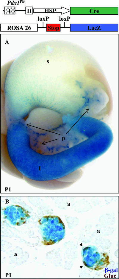
The critical pancreatic transcription factor Pdx1 is expressed throughout the pancreas early but enriched in insulin-producing beta cells postnatally. Previous studies showed that the 5' conserved promoter regions areas I and II (Pdx1(PB)) direct endocrine cell expression, while an adjacent region (Pdx1(XB)) containing conserved area III directs transient beta-cell expression. In this study, we used Cre-mediated lineage tracing to track cells that activated these regions. Pdx1(PB)Cre mediated only endocrine cell recombination, while Pdx1(XB)Cre directed broad and early recombination in the developing pancreas. Also, a reporter transgene containing areas I, II, and III was expressed throughout the embryonic day 10.5 (E10.5) pancreas and gradually became beta cell enriched, similar to endogenous Pdx1. These data suggested that sequences within area III mediate early pancreas-wide Pdx1 expression. Area III contains a binding site for PTF1, a transcription factor complex essential for pancreas development. This site contributed to area III-dependent reporter gene expression in the acinar AR42J cell line, while PTF1 specifically trans-activated area III-containing reporter expression in a nonpancreatic cell line. Importantly, Ptf1a occupied sequences spanning the endogenous PTF1 site in area III of E11.5 pancreatic buds. These data strongly suggest that PTF1 is an important early activator of Pdx1 in acinar and endocrine progenitor cells during pancreas development.


Figures

FIG. 1.
FIG. 1.
Diagram of highly conserved areas within the Pdx1 promoter/enhancer region. Area I (bp −2852 to −2547), area II (bp −2247 to −2071), area III (bp −1973 to −1694), and area IV (bp −6422 to −5931) are conserved regions among species (14). Labeled horizontal lines below the Pdx1 promoter indicate regions of relevance in this investigation. PstI (−3007), XhoI (−2046), BstEII (−2011), and BglII (−994) are relevant restriction sites. The numbering is relative to the mouse Pdx1 gene translation start site.
FIG. 2.
FIG. 2.
Lineage tracing of a region that contains the conserved area I and II (Pdx1PBCre)-mediated recombination only in islet endocrine cells of the pancreas at P1. (A) Whole mount of X-Gal-stained (blue) P1 digestive organs from a Pdx1PBCre; R26R mouse showing high levels of expression in islets and some cells within the stomach. The arrows indicate different regions of the pancreas containing X-Gal-stained cells consistent with normal islet distribution. X-Gal staining was sometimes observed in the duodenal lumen of transgenic and nontransgenic neonatal littermates. p, pancreas; s, stomach; I, intestine. (B) Cross-section of X-Gal-stained P1 pancreas tissue labeled with antibodies to glucagon (Gluc) in brown. The arrowheads point to cells positive for both glucagon and β-Gal. a, acinar tissue. The diagram shows a schematic of the Pdx1PBCre transgene and R26R reporter construct for lineage tracing of Cre-expressing cells (not to scale).
FIG. 3.
FIG. 3.
Expression is restricted to β cells in Pdx1I-II-IIIlacZ adults. (A, B) Whole-mount pictures of X-Gal-stained (blue) adult pancreata from two independent transgenic lines showing an islet-enriched expression pattern. (C, D) Representative cross-sections of X-Gal-stained adult pancreata, counterstained with eosin, which was typically detected throughout the β cells (C), but sometimes appeared variegated (D). Glucagon-expressing cells are labeled with an antibody in brown (Gluc). The original magnification is given for each panel. The diagram refers to the transgene used to create the mice analyzed in this figure (not to scale).
FIG. 4.
FIG. 4.
Pdx1I-II-IIIlacZ is expressed in both endocrine and acinar cells during development. (A, B) X-Gal-stained (blue) embryos at E11.5 showing expression in pancreas and ectopic expression in the neural tube. (C) Section from an X-Gal-stained E10.5 embryo labeled for Pdx1 protein (brown). (D, E) X-Gal staining in the developing pancreas at E10.5 (D) and E11.5 (E). (F, G, H) Whole-mount pictures of X-Gal-stained dissected digestive organs at E16.5 (F), E18.5 (G), and P1 (H). The pictures are representative of expression observed in four of four independent transgenic mice. Luminal X-Gal staining was observed in the duodenum of some transgenic and nontransgenic littermates at late gestation and in neonates (G). p, pancreas; sp, spleen; s, stomach; I, rostral duodenal portion of the intestine; db, pancreatic dorsal bud; vb, ventral bud. The diagram refers to the transgene used to create the mice analyzed in this figure (not to scale).
FIG. 5.
FIG. 5.
Lineage tracing of area III-containing, Pdx1XBCre-mediated recombination throughout the endogenous Pdx1 domain. Whole-mount pictures of X-Gal-stained E10.5 (B) and E11.5 (E) embryos and dissected digestive organs at E10.5 (A), E11.5 (D), E14.5 (F), and P1 (G). (C) Cross-section of X-Gal-stained P1 pancreas labeled with antibodies to glucagon (Gluc; brown). The pictures are representative of expression observed in four of four independent transgenic mice. L, liver; s, stomach; I, rostral duodenal portion of the intestine; b, bile duct; i, pancreatic islet; a, acinus; d, duct; db, dorsal bud; vb, ventral bud. The diagram refers to the transgene used to create the mice analyzed in this figure and the R26R reporter construct for lineage tracing of Cre-expressing cells (not to scale).
FIG. 6.
FIG. 6.
Ptf1a-containing complex binds area III and requires both the E and TC boxes. (A) Gel shift using the putative PTF1 binding site from area III or the PTF1 binding site from the elastase (Ela1) promoter as a control (closed arrowhead refers to specific complex). The addition of Ptf1a antibody (Ab) results in a complete supershift of the PTF1 complex bound to both the AIII probe and Ela1 PTF1 control probe (open arrowhead). The triangles depict increasing amounts of cold competitor: 10-, 50-, and 100-fold. The rectangle represents 100-fold competitor. Panc nuc, pancreatic nuclear extract; Min6 nuc, MIN-6 nuclear extract. (B) Gel shift competition experiment using indicated E box- or TC box-mutant cold competitors. The triangle depicts increasing amounts of cold competitor: 10-, 100-, and 1,000-fold. The rectangles represent 1,000-fold cold competitor. (C) Sequences of oligonucleotides used in the gel shift experiments. Dashes indicate gaps, periods indicate homology to AIII PTF1 probe, and capital letters indicate conserved nucleotides. Dup, duplication of spacer sequence.
FIG. 7.
FIG. 7.
Pdx1 area III activity is dependent on PTF1. (A) Wild-type (wt) and mutated (mutant) PTF1 binding site TK-Luc reporters were transfected into AR42J cells. Activity is expressed as percent Pdx1I-II-III activation ± standard error of the mean. (B) Expression plasmids of Ptf1a, HEB, and RBP-Jκ or -L were cotransfected into HEK 293 cells with the area III-containing TK-Luc plasmids. The data are expressed as fold activation ± standard error of the mean over the respective area III-containing TK-Luc cotransfected with empty expression plasmids. The asterisks indicate significance upon comparing the mutant to the wild type: *, P < 0.01, **, P < 0.001. The diagrams at the bottom of each panel refer to enhancer fragments used to drive reporter expression.
FIG. 8.
FIG. 8.
Ptf1a protein is specifically enriched at area III and area IV of the Pdx1 promoter. (A to F) The midgut region (A, B) was isolated from E11.5 embryos. The region containing the dorsal pancreas was removed (C, D) and separated from contaminating tissue (E). Several dissected pancreatic buds were pooled and processed for chromatin cross-linking and chromatin isolation at one time (F). (G) Tissue purity was assayed by determining Ptf1a or alb1 expression level. mRNA isolated from dissected organs was subjected to real-time reverse transcriptase-PCR analysis and normalized to hypoxanthine phosphoribosyltransferase (HPRT) mRNA (see Materials and Methods). (H) Ptf1a ChIP was performed on chromatin isolated from E11.5 dorsal pancreas and gut tube. Detection of enrichment for the elastase 1 promoter serves as a positive control for Ptf1a binding and failure of Ptf1a to precipitate chromatin from the gut tube demonstrates tissue specificity. The data represent the averages of two Bioanalyzer runs per primer pair set amplified in parallel from single ChIP reactions (IgG or Ptf1a) from designated embryonic tissues. The diagram refers to the location of the amplified regions in relation to the transcription start site.

References

    1. Ahlgren, U., J. Jonsson, L. Jonsson, K. Simu, and H. Edlund. 1998. Beta-cell-specific inactivation of the mouse Ipf1/Pdx1 gene results in loss of the beta-cell phenotype and maturity onset diabetes. Genes Dev. 12:1763-1768. - PMC - PubMed
    1. Beres, T. M., T. Masui, G. H. Swift, L. Shi, R. M. Henke, and R. J. MacDonald. 2006. PTF1 is an organ-specific and Notch-independent basic helix-loop-helix complex containing the mammalian suppressor of hairless (RBP-J) or its paralogue, RBP-L. Mol. Cell. Biol. 26:117-130. - PMC - PubMed
    1. Bossard, P., and K. S. Zaret. 2000. Repressive and restrictive mesodermal interactions with gut endoderm: possible relation to Meckel's diverticulum. Development 127:4915-4923. - PubMed
    1. Boyer, D. F., Y. Fujitani, M. Gannon, A. C. Powers, R. W. Stein, and C. V. Wright. 2006. Complementation rescue of Pdx1 null phenotype demonstrates distinct roles of proximal and distal cis-regulatory sequences in pancreatic and duodenal expression. Dev. Biol. 298:616-631. - PubMed
    1. Brissova, M., M. Shiota, W. E. Nicholson, M. Gannon, S. M. Knobel, D. W. Piston, C. V. Wright, and A. C. Powers. 2002. Reduction in pancreatic transcription factor PDX-1 impairs glucose-stimulated insulin secretion. J. Biol. Chem. 277:11225-11232. - PubMed

Publication types

MeSH terms

Substances