. 2007 Jun;27(11):4070-81.
doi: 10.1128/MCB.02011-06. Epub 2007 Apr 2.

RIAM links the ADAP/SKAP-55 signaling module to Rap1, facilitating T-cell-receptor-mediated integrin activation

Affiliations

RIAM links the ADAP/SKAP-55 signaling module to Rap1, facilitating T-cell-receptor-mediated integrin activation

Gaël Ménasché et al. Mol Cell Biol. 2007 Jun.

Abstract

One outcome of T-cell receptor (TCR) signaling is increased affinity and avidity of integrins for their ligands. This occurs through a process known as inside-out signaling, which has been shown to require several molecular components including the adapter proteins ADAP (adhesion and degranulation-promoting adapter protein) and SKAP-55 (55-kDa src kinase-associated phosphoprotein) and the small GTPase Rap1. Herein, we provide evidence linking ADAP and SKAP-55 to RIAM, a recently described adapter protein that binds selectively to active Rap1. We identified RIAM as a key component linking the ADAP/SKAP-55 module to the small GTPase Rap1, facilitating TCR-mediated integrin activation. We show that RIAM constitutively interacts with SKAP-55 in both a heterologous transfection system and primary T cells and map the region essential for this interaction. Additionally, we find that the SKAP-55/RIAM complex is essential both for TCR-mediated adhesion and for efficient conjugate formation between T cells and antigen-presenting cells. Mechanistic studies revealed that the ADAP/SKAP-55 module relocalized RIAM and Rap1 to the plasma membrane following TCR activation to facilitate integrin activation. These results describe for the first time a link between ADAP/SKAP-55 and the Rap1/RIAM complex and provide a potential new mechanism for TCR-mediated integrin activation.


Figures
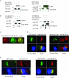
FIG. 1.
FIG. 1.
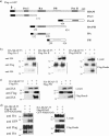
The adapter protein RIAM interacts with SKAP-55 in transfected 293T cells and colocalizes with SKAP-55 in the Jurkat T-cell line. (a) (Left) His-ADAP and Flag-RIAM were cotransfected into 293T cells. Cell lysates were immunoprecipitated with anti-Flag and then separated by SDS-polyacrylamide gel electrophoresis. Coprecipitated RIAM and ADAP were examined by immunoblot analysis with anti-His and anti-Flag. (Right) Flag-ADAP and GFP-RIAM were cotransfected into 293T cells. Cell lysates were immunoprecipitated with anti-Flag and then separated by SDS-polyacrylamide gel electrophoresis. Coprecipitated RIAM and ADAP were examined by immunoblot analysis with anti-GFP and anti-Flag. (b) (Left) HA-SKAP-55 and Flag-RIAM were cotransfected into 293T cells. Cell lysates were immunoprecipitated with anti-Flag. Coprecipitated RIAM and SKAP-55 were examined by immunoblot analysis with anti-HA and anti-Flag. (Right) Flag-SKAP-55 and GFP-RIAM or Flag-SLP-76 and GFP-RIAM were cotransfected into 293T cells. The anti-Flag immunoprecipitation reveals only an interaction between SKAP-55 and RIAM. (c) Jurkat T cells were cotransfected with GFP-RIAM and HA-SKAP-55 constructs. Fixed cells were labeled with an anti-HA antibody followed by anti-rat antibody-Alexa Fluor 594 staining (red). (d) The Jurkat T cells transfected with either GFP-RIAM or HA-SKAP-55 were used to form conjugates with SEE-pulsed Raji B cells. The Raji cells were also labeled in blue with the cell tracker Blue CMAC. Cells were then fixed, permeabilized, and stained with phalloidin 594 (red) or phalloidin 488 (green). Conjugates were analyzed for accumulation of HA-SKAP-55 or GFP-RIAM at the T-B interface. (e) Jurkat T cells were cotransfected with GFP-RIAM and HA-SKAP-55. Conjugates were formed and analyzed for HA-SKAP-55 and GFP-RIAM colocalization. Representative conjugates are shown. Each study was repeated at least three times, and in each study, more than 30 conjugates were examined per condition.
FIG. 2.
FIG. 2.
Mapping the RIAM domains involved in SKAP-55 binding. (a) Schematic representation of the RIAM constructs used in these experiments. (b) Each domain of RIAM fused to a Flag tag was cotransfected with HA-SKAP-55 in 293T cells. Cell lysates were immunoprecipitated with anti-Flag and then separated by SDS-polyacrylamide gel electrophoresis. Coprecipitated RIAM domains and SKAP-55 were examined by immunoblot analysis with anti-HA and anti-Flag. (c) Flag-RIAM RA or Flag-RIAM PH was cotransfected along with HA-SKAP-55. Cell lysates were immunoprecipitated with anti-Flag. Coprecipitated domains of RIAM and SKAP-55 were examined by immunoblot analysis with anti-HA and anti-Flag. (d) The HA-SKAP-55 and Flag-RIAM were cotransfected with the GFP-RIAM RA or GFP-RIAM PH domain in 293T cells.
FIG. 3.
FIG. 3.
The SKAP-55/RIAM complex interacts with ADAP and the active form of Rap1. (a) Flag-ADAP, HA-SKAP-55, and GFP-RIAM were transfected in 293T cells. Cell lysates were immunoprecipitated with anti-Flag and then examined by immunoblot analysis using anti-GFP, anti-HA, and anti-Flag. (b) GFP-Rap1Q63E, HA-SKAP-55, and Flag-RIAM were transfected in 293T cells. Cell lysates were immunoprecipitated with anti-Flag. RIAM coprecipitates were examined by immunoblot analysis with anti-GFP, anti-HA, and anti-Flag. (c) Cell lysates containing GFP-Rap1Q63E, Flag-RIAM, and GFP-RIAM RA domain were immunoprecipitated with anti-Flag. The immunoblots were revealed using anti-GFP and anti-Flag. (d) Cell lysates containing GFP-Rap1Q63E, Flag-RIAM, and GFP-RIAM PH domain were immunoprecipitated with anti-Flag. The immunoblots were analyzed using anti-GFP and anti-Flag.
FIG. 4.
FIG. 4.
RIAM immunoprecipitates SKAP-55 and ADAP in primary T cells. (a) Murine T-cell blasts were rested or activated by cross-linking the TCR with an anti-CD3 antibody or with the phosphatase inhibitor PV. Cell lysates were immunoprecipitated with an isotype control or with an anti-RIAM polyclonal antibody and immunoblotted with anti-RIAM, anti-SKAP-55, and anti-ADAP. Cell lysates were also immunoblotted for the presence of ERK and phospho-ERK. (b) Murine T-cell BLAST lysates were immunoprecipitated with anti-RIAM or anti-ADAP. ADAP immunoprecipitations (one for lane 5 and two for lane 6) were performed before the RIAM immunoprecipitation. The immunoblots were analyzed using anti-RIAM, anti-SKAP-55, and anti-ADAP. (c) Jurkat T cells or primary human T cells (hT-cells) were stimulated for 5 min with MAb to CD3 (+) (C305 or MEM92) or left untreated (−). Cells were lysed, and immunoprecipitations were performed using anti-ADAP or an isotype control. Precipitates were analyzed by Western blot analysis for the presence of ADAP, SKAP-55, RIAM, or Rap1. Successful stimulation of T cells was assessed by analyzing the phosphorylation status of ERK. IP, immunoprecipitation; P-ERK, phospho-ERK; Iso, isotype control.
FIG. 5.
FIG. 5.
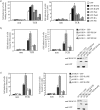
Disruption of the SKAP-55/RIAM complex interferes with the TCR-dependent β1- and β2-integrin activation. (a) Jurkat T cells were transfected with cDNAs encoding GFP, GFP-RIAM, GFP-RAPH, GFP-ProI, or GFP-ProII. Transfectants either were left untreated (non) or were stimulated with anti-CD3 OKT3 (TCR) for 30 min and subsequently analyzed for their ability to adhere to fibronectin or to ICAM-1. Adhesion data represent the means ± the standard errors of the means of three independently performed experiments. (b) Jurkat T cells transfected with pS-SK1 (siRNA directed against SKAP-55) or pS-SK4 (scrambled siRNA) were cotransfected with either GFP alone or GFP-RIAM and analyzed for adhesion to fibronectin or ICAM-1 under resting (non) or TCR-stimulated (TCR) conditions. (c) Jurkat T cells transfected with pS-SK1 or pS-SK4 were transfected with GFP or GFP-RAPH and then analyzed for adhesion to fibronectin or ICAM-1 under resting (non) or TCR-stimulated (TCR) conditions.
FIG. 6.
FIG. 6.
Impaired conjugate formation between B cells and Jurkat cells expressing the RIAM RA or RIAM PH domain. Jurkat T cells transiently transfected with either GFP, GFP-RA, or GFP-PH were incubated with unpulsed or SEE-pulsed DDAO-SE-labeled Raji B cells. The percentage of conjugate formation between Jurkat T cells and Raji B cells was determined by FACS analysis. A histogram representative of a set of two-dimensional plots is shown. Data are the average means and standard errors of the means from three independent experiments.
FIG. 7.
FIG. 7.
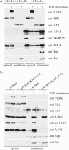
Disruption of the ADAP/SKAP-55 module interferes with the plasma membrane localization of RIAM and Rap1. (a) Purified splenic T cells prepared from ADAP-deficient [ADAP (−/−)] or wild-type (wt) mice were preincubated with biotinylated anti-CD3 MAb (2C11) and then cross-linked with streptavidin for 3 min at 37°C. Subsequently, membrane and cytosolic fractions were prepared from unstimulated (−) or stimulated (+) cells. The individual samples were analyzed by Western blotting using anti-ADAP, anti-SKAP-55, anti-RIAM, anti-Rap1, and anti-Ras. To control the fractionation efficiency and proper TCR stimulation, fractions were assessed for the presence of LAT (plasma membrane), ERK (cytosol), or phospho-ERK (stimulation) (P-ERK). (b) Jurkat T cells were transiently transfected with either pS-SK4 (scrambled) or pS-SK1 (siRNA of SKAP-55) or were either left unstimulated (−) or stimulated for 5 min with the anti-TCR MAb C305. Cytosolic and membrane fractions were analyzed as described above.
FIG. 8.
FIG. 8.
Disruption of the ADAP/SKAP-55 module interferes with plasma membrane localization of both Rap1G12V and RIAM. (a) Jurkat T cells were transfected with constructs encoding a scrambled RNAi or an RNAi directed against SKAP-55 along with a construct encoding DsRed-tagged Rap1G12V. Cells were stimulated for 5 min through the TCR and were then imaged by confocal laser scanning microscopy. Rap1G12V translocation to the membrane was analyzed by determining the intensity of DsRed staining using Corel software (60 cells examined for each experiment). The percentage of total pixels representing Rap1G12V at the membrane for each cell is shown in the scattergram (left). This study is representative of three independent experiments. In parallel, transfectants were analyzed for their capability to adhere to fibronectin. The adhesion results are shown in the bar graph (right) and represent the means ± the standard errors of the means of three independent experiments. (b) Purified splenic T cells prepared from ADAP-deficient [ADAP (−/−)] or wild-type mice were preincubated with biotinylated anti-CD3 MAb and then cross-linked with streptavidin for 3 min at 37°C. Cells were fixed, permeabilized, and stained with phalloidin-tetramethyl rhodamine isocyanate (TRITC) (red) and RIAM in combination with anti-rabbit IgG-fluorescein isothiocyanate.

Similar articles

Cited by

References

    1. Bazzoni, G., and M. E. Hemler. 1998. Are changes in integrin affinity and conformation overemphasized? Trends Biochem. Sci. 23:30-34. - PubMed
    1. Bivona, T. G., H. H. Wiener, I. M. Ahearn, J. Silletti, V. K. Chiu, and M. R. Philips. 2004. Rap1 up-regulation and activation on plasma membrane regulates T cell adhesion. J. Cell Biol. 164:461-470. - PMC - PubMed
    1. Boerth, N. J., B. A. Judd, and G. A. Koretzky. 2000. Functional association between SLAP-130 and SLP-76 in Jurkat T cells. J. Biol. Chem. 275:5143-51452. - PubMed
    1. Bos, J. L. 2005. Linking Rap to cell adhesion. Curr. Opin. Cell Biol. 17:123-128. - PubMed
    1. Bos, J. L., B. Franke, L. M'Rabet, K. Reedquist, and F. Zwartkruis. 1997. In search of a function for the Ras-like GTPase Rap1. FEBS Lett. 410:59-62. - PubMed

Publication types

MeSH terms