. 2007 Apr 9;177(1):13-20.
doi: 10.1083/jcb.200612140. Epub 2007 Apr 2.

A role for a novel centrosome cycle in asymmetric cell division

Affiliations

A role for a novel centrosome cycle in asymmetric cell division

Nasser M Rusan et al. J Cell Biol. .

Abstract

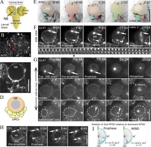
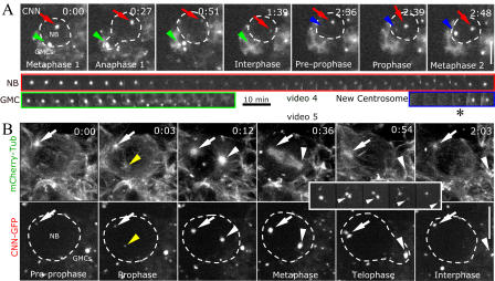
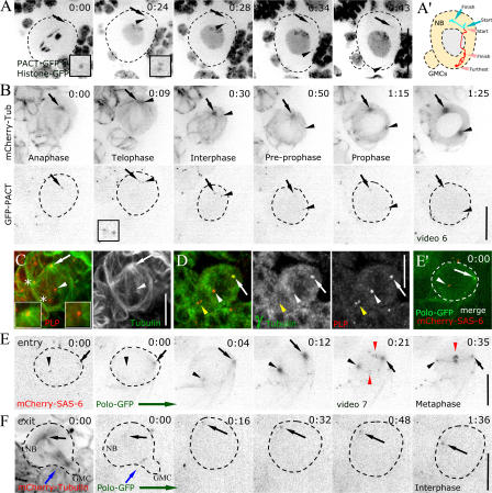
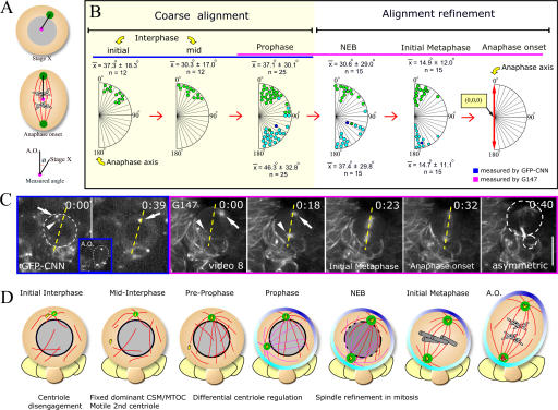
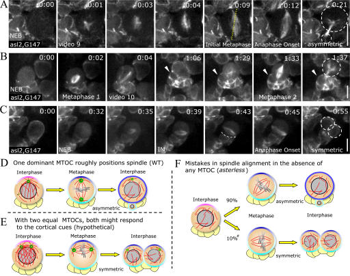
Tissue stem cells play a key role in tissue maintenance. Drosophila melanogaster central brain neuroblasts are excellent models for stem cell asymmetric division. Earlier work showed that their mitotic spindle orientation is established before spindle formation. We investigated the mechanism by which this occurs, revealing a novel centrosome cycle. In interphase, the two centrioles separate, but only one is active, retaining pericentriolar material and forming a "dominant centrosome." This centrosome acts as a microtubule organizing center (MTOC) and remains stationary, forming one pole of the future spindle. The second centriole is inactive and moves to the opposite side of the cell before being activated as a centrosome/MTOC. This is accompanied by asymmetric localization of Polo kinase, a key centrosome regulator. Disruption of centrosomes disrupts the high fidelity of asymmetric division. We propose a two-step mechanism to ensure faithful spindle positioning: the novel centrosome cycle produces a single interphase MTOC, coarsely aligning the spindle, and spindle-cortex interactions refine this alignment.


Figures

Figure 1.
Figure 1.
NB MTOCs form asynchronously. (A) Cartoon. (B–E) NB/GMCs in central brain (B), close-up (C and E), and cartoon (D). (B and C) Phalloidin. (E) Actin-GFP for two cell cycles; positions of successive GMCs are indicated (colored). The yellow dot represents a hypothetical MTOC. (F and G) GFP-G147 NBs. (F) GMCs are indicated by the dotted line. Max-intensity projections are shown for entire cell. Single MTOC matures and forms MT basket (0:00; arrow). Second MTOC appears on other side of nucleus (1:45; arrowhead) and matures (4:35). Small images show dominant MTOC (top) and second MTOC (bottom; asterisk). (G) An end-on view of MTOC maturation, three sections of z stack. Dominant MTOC is present throughout (top, arrows). Second MTOC appears (7:74; arrow). Dotted line indicates bottom of NB. (H) EB1-GFP. Second nucleation center appears (1:09; arrow). (I) Position where second MTOC appears relative to dominant MTOC. Time is shown as h:min (E) and min:s (F–H). Bars, 10 μm.
Figure 2.
Figure 2.
Differential maturation of NB centrosomes. (A) GFP-Cnn for one cell cycle. Dominant centrosome is indicated by the red arrows. PCM is reduced during mitotic exit and accumulates in preprophase. GMC centrosome completely loses PCM (green arrowheads). Second centrosome appears distant from dominant centrosome (blue arrowheads). The asterisk highlights the appearance of the second centrosome. (B) GFP-Cnn, chTub. Arrows indicate dominant centrosome. Arrowheads indicate second centrosome. The inset shows GFP-Cnn, PCM splitting. Time is shown as h:min. Bars, 10 μm.
Figure 3.
Figure 3.
NB centrioles differ in behavior and regulation. NBs are outlined in A, B, E, and F. Imaged proteins are indicated on each figure. Images are displayed in inverse contrast. (A) Centrioles separate (each indicated by an arrow or arrowhead; 0:24; insets highlight the separation). One is relatively stationary (arrows), and the second moves to other side of nucleus (arrowheads). (A′) The diagram shows the path of centrioles in A. (B) Centrioles split in late telophase; one remains stationary with associated MTs (arrows), and the other loses MTs and moves around the nucleus (arrowheads). (C and D) Fixed NBs. DPLP and MTs (C) or γtub (D) are shown. (C) Preprophase NB. One centriole is at the center of MT aster (arrows), and the other has no associated MTs (arrowheads). Asterisks show centrioles in adjacent cells that appear to be in NB by max-intensity projection. (D) Only the centriole distant from GMC cap has γtub (arrows). GMC centrioles lack γtub (yellow arrowheads). (E and E′) Mitotic entry: preprophase dominant centriole has Polo (arrows), and the second does not (arrowheads). (0:04) Prophase; second centriole begins to acquire Polo (arrowheads). After NEB, Polo moves to kinetochores (0:21–0:35; red arrowheads). (F) Mitotic exit: NB retains Polo on dominant centriole from telophase into interphase (0:00–1:36; black arrows). Polo is also at midbody in telophase (blue arrows). Time is shown as h:min. Bars, 10 μm.
Figure 4.
Figure 4.
Two phases of spindle alignment. (A) Cartoon showing measured angles. (B) Centrosome location relative to anaphase division axis onset. Green circles indicate the dominant centrosome, and blue circles indicate the second centrosome. In 1/25 NBs, the dominant centrosome (dark green) was on the GMC side of the nucleus at prophase; its second centrosome is shown in dark blue. Measurements used Cnn (blue; interphase and prophase) or MTs (pink). (C) Sample video stills. Yellow lines indicate anaphase-onset axis. (D) Cartoon showing centrosome/centriole cycles. Time is shown as h:min. Bar, 10 μm.
Figure 5.
Figure 5.
Functional centrosomes ensure high-fidelity division asymmetry. (A–C) GFP-G147 in asl. (A) Chromosome-induced spindle assembly (0:01–0:09). Initial spindle alignment is absent, but refinement occurs (0:09–0:12). (B) Two rounds of mitosis. GMCs born near one another. Arrowheads indicate first daughter. (C) Example where initial spindle alignment was far off NB-GMC axis, with resulting symmetric division. (D) Mechanistic model of the importance of dominant interphase MTOC. (E) Hypothetical case: centrosomes matured synchronously as in canonical cycle. (F) Division with no centrosomes. Pound sign indicates that number is from fixed analysis of telophase NBs (Giansanti et al., 2001). Time is shown as h:min. Bars, 10 μm.

References

    1. Akong, K., B.M. McCartney, and M. Peifer. 2002. Drosophila APC2 and APC1 have overlapping roles in the larval brain despite their distinct intracellular localizations. Dev. Biol. 250:71–90. - PubMed
    1. Alberts, B., A. Johnson, J. Lewis, M. Raff, K. Roberts, and P. Walter. 2002. Molecular biology of the cell. Garland Science, New York. 1616 pp.
    1. Basto, R., J. Lau, T. Vinogradova, A. Gardiol, C.G. Woods, A. Khodjakov, and J.W. Raff. 2006. Flies without centrioles. Cell. 125:1375–1386. - PubMed
    1. Callaini, G., and M.G. Riparbelli. 1990. Centriole and centrosome cycle in the early Drosophila embryo. J. Cell Sci. 97:539–543. - PubMed
    1. Ceron, J., C. Gonzalez, and F.J. Tejedor. 2001. Patterns of cell division and expression of asymmetric cell fate determinants in postembryonic neuroblast lineages of Drosophila. Dev. Biol. 230:125–138. - PubMed

Publication types

MeSH terms

Substances