. 2007 Apr 9;177(1):151-62.
doi: 10.1083/jcb.200701086. Epub 2007 Apr 2.

c-Met is essential for wound healing in the skin

Affiliations

c-Met is essential for wound healing in the skin

Jolanta Chmielowiec et al. J Cell Biol. .

Abstract

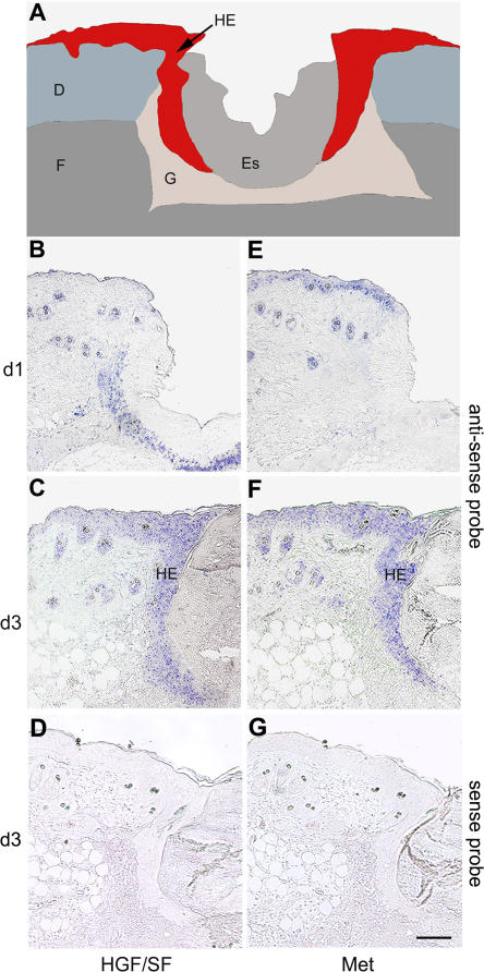
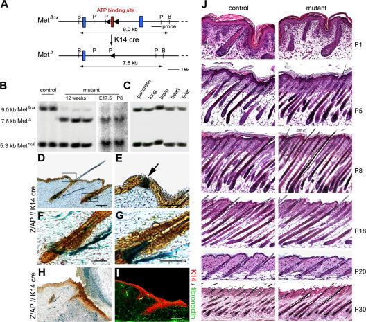
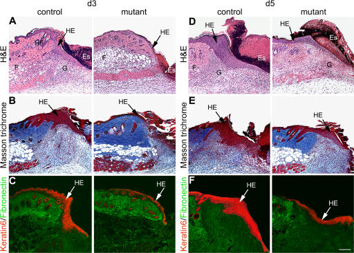
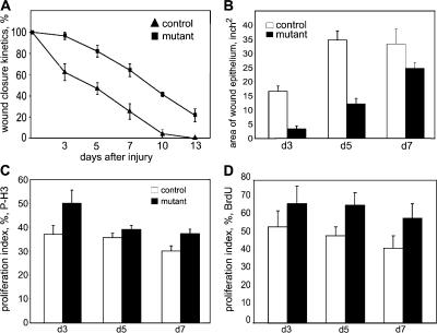
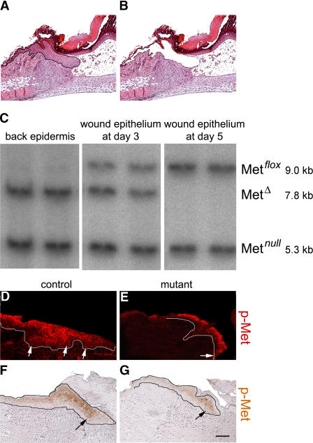
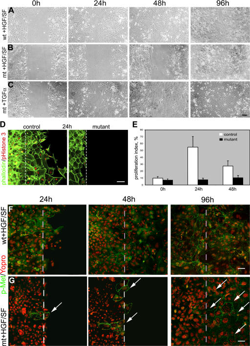
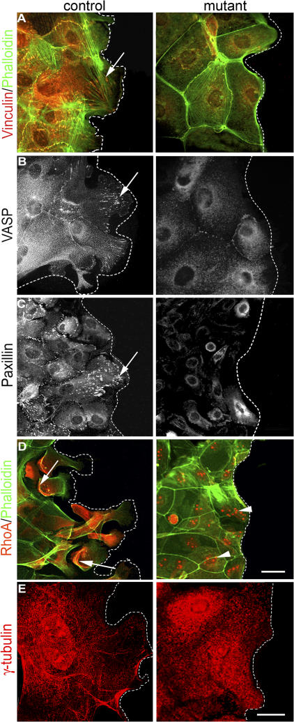
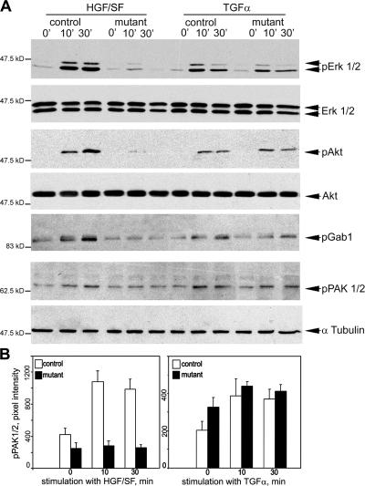
Wound healing of the skin is a crucial regenerative process in adult mammals. We examined wound healing in conditional mutant mice, in which the c-Met gene that encodes the receptor of hepatocyte growth factor/scatter factor was mutated in the epidermis by cre recombinase. c-Met-deficient keratinocytes were unable to contribute to the reepithelialization of skin wounds. In conditional c-Met mutant mice, wound closure was slightly attenuated, but occurred exclusively by a few (5%) keratinocytes that had escaped recombination. This demonstrates that the wound process selected and amplified residual cells that express a functional c-Met receptor. We also cultured primary keratinocytes from the skin of conditional c-Met mutant mice and examined them in scratch wound assays. Again, closure of scratch wounds occurred by the few remaining c-Met-positive cells. Our data show that c-Met signaling not only controls cell growth and migration during embryogenesis but is also essential for the generation of the hyperproliferative epithelium in skin wounds, and thus for a fundamental regenerative process in the adult.


Figures

Figure 1.
Figure 1.
Expression of HGF/SF and c-Met during wound healing. (A) Scheme of a wound section 3–5 d after injury. Keratinocytes (red) at the wound edge proliferate and migrate down the injured dermis to form the so-called HE (arrow). D, dermis; Es, eschar; F, fatty tissue; G, granulation tissue. (B and C) In situ hybridization of wounded skin with a HGF/SF probe 1 (B) and 3 d (C) after injury. Note that HGF/SF at day 1 is expressed in the hair follicles and around the clot, which is where inflammatory cells accumulate and infiltrate the wound. 3 d after wounding, HGF/SF is highly expressed in the HE. (E and F) In situ hybridization with the c-Met probe 1 (D) and 3 d (E) after injury. Note that c-Met is expressed in the epidermis, in hair follicles, and in the HE 3 d after wounding. (D and G) In situ hybridization with sense probes of HGF/SF (F) and c-Met (G). Bar, 50 μm.
Figure 2.
Figure 2.
Generation and analysis of skin-specific c-Met mutant mice. (A) Schematic representation of nonrecombined allele and recombined allele of c-Met. Exon 15 of the c-Met gene that encodes the ATP-binding site (red box) was flanked by loxP sites (triangles) and is excised after K14-cre–induced recombination. Blue boxes indicate exons 14 and 16. The sizes of the restriction fragments generated by BamHI digest before and after recombination are indicated. B, BamHI; P, Pst. (B) Southern blot analysis of epidermis from control and conditional c-Met mutant mice of different ages (12 wk old, E17.5, and P8). (C) Southern blot analysis of different organs of conditional c-Met mutant mice. (D–G) Double staining of skin section from Z/AP; K14-cre mice (Lobe et al., 1999; Huelsken et al., 2001) for lacZ (blue, nonrecombined) and alkaline phosphatase activity (yellow, recombined). (E) A higher magnification shows an area of nonrecombined epidermal keratinocytes (blue patch). Higher magnifications show also two independent hair follicles (F and G); note that the bulge region contains recombined (yellow) cells. Arrector pili muscle cells, which surround the follicle, are nonrecombined (blue). (H) Double staining of alkaline phosphatase and β-galactosidase activity of wound section from Z/AP; K14-cre mice. Note that K14-cre–induced recombination is observed in the unwounded epidermis and in the HE. (I) Immunohistological analysis of a wound section from control mice using antibodies directed against K14 (red) and fibronectin (green). (J) Hair follicle cycle in control and conditional c-Met mutant. Sagittal sections of control and conditional c-Met mutant skin stained with hematoxylin/eosin at P1 (first anagen), P5 (first anagen), P8 (first anagen), P18 (first catagen), P20 (first telogen), and P30 (second anagen). Bars: (D) 50 μm; (E–F) 20 μm; (G–I) 100 μm.
Figure 3.
Figure 3.
Wound healing in control and conditional c-Met mutant mice. (A and D) Hematoxylin/eosin staining of sections of wound edges from control and mutant mice 3 (A) and 5 d (D) after wounding. Only halves of the wounds are shown (for the scheme of a complete wound see Fig. 1). (B and E) Masson trichrome staining of wound sections 3 (B) and 5 d (E) after injury. (C and F) Immunohistological analysis of wound sections from control and mutant mice 3 (C) and 5 d (F) after injury using antibodies directed against keratin 6 (red) and fibronectin (green). Arrows mark the HE. Es, eschar; F, fatty tissue; G, granulation tissue; HF, hair follicle. Bar, 100 μm.
Figure 4.
Figure 4.
Quantification of wound healing in control and conditional c-Met mutant mice. (A) Wound closure kinetics in control and mutant mice (see Materials and methods). (B) Quantification of the area of HE 3, 5, and 7 d after wounding in control and mutant mice; only sections of the middle of the wounds were used for quantification. (C) Proliferation of keratinocytes in the HE from control and mutant mice 3, 5, and 7 d after wounding, as assessed by the proportion of phospho-Histone 3–positive nuclei in the epithelium (see Materials and methods). A t test was performed, and significant differences between control and mutant were observed 3 d after injury. P = 0.01. (D) Proliferation of keratinocytes in the HE from control and mutant mice 3, 5, and 7 d after wounding, as assessed by the proportion of BrdU-positive nuclei in the epithelium. Significant statistical differences between control and mutant were observed 5 d after injury. P = 0.01. Error bars represent the SD.
Figure 5.
Figure 5.
Only residual c-Met–positive keratinocytes contribute to the HE of wounds in conditional c-Met mutant mice. (A and B) Isolation of HE by laser capture microdissection. A wound section before (A) and after laser capture microdissection (B) is shown. (C) Southern blot analyses of back epidermis and hyperproliferative epithelia from conditional c-Met mutant mice. Microdissected, nonwounded epidermis from the back (left) and microdissected hyperproliferative epithelia of wounds 3 (middle) and 5 d (right) after injury were collected. Southern blotting of two independent preparations from different pools of microdissected tissues is shown. Note that the HE 5 d after injury in conditional c-Met mutant mice is formed exclusively by cells that contain the nonrecombined c-Metflox allele, but not the recombined c-MetΔ allele. At day 3, a 1:1 mixture of recombined and nonrecombined cells is seen (middle). The c-Metnull allele is also present because heterozygous c-Metflox/null mice were used for conditional mutagenesis. (D–G) Immunohistological analysis of wound sections from control and conditional mutant mice 5 d after injury using anti–phospho-c-Met antibodies (red immunofluorescence in D and E, and brown immunohistochemistry in F and G). Note that in the skin of conditional c-Met mutant mice, cells in the HE (outlined) are phospho-c-Met positive. Arrows mark phospho-c-Met–positive cells in the lower hyperproliferative epithelia layers. Bar, 100 μm.
Figure 6.
Figure 6.
Scratch wound healing in cell culture of primary keratinocytes: c-Met–positive primary keratinocytes migrate preferentially into scratch wounds. Primary keratinocytes were isolated from newborn skin of control (A) and conditional c-Met mutant mice (B and C). After scratch wounding, cells were further cultured in the presence of HGF/SF or TGFα. Photos were taken 0, 24, 48, and 96 h after scratch wounding. Note that wounds in the cultures derived from conditional mutant mice did only close after 96 h in the presence of HGF/SF. (D) Proliferation of primary keratinocytes from control and conditional c-Met mutant mice 24 h after stimulation with HGF/SF, as assessed by phospho-histone 3 antibody staining (red). A dashed line marks the scratch edge. Counterstaining was performed with phalloidin (green). (E) Quantification of proliferation of primary keratinocytes at wound edges stimulated with HGF/SF in the experiments described in D. Error bars represent the SD. (F and G) Primary keratinocytes isolated from control and conditional c-Met mutant skin were scratch wounded and further cultured with HGF/SF. After 24, 48, and 96 h, cells were stained with anti–phospho-c-Met antibodies (green). Nuclei were visualized by YO-PRO staining (red). Note that many cells in the controls (F) showed a cobblestone pattern of phospho-c-Met staining at the plasma membrane. In populations derived from the skin of conditional mutant mice (G), cells with phospho-c-Met were initially rare (at 24 h), but after 96 h they are abundant in the scratched area (marked by arrows). The original edges of the scratch wounds are marked with a dashed line. Bars: (A–D) 100 μm; (F and G) 50 μm.
Figure 7.
Figure 7.
Keratinocytes derived from conditional c-Met mutant mice do not rearrange their cytoskeleton at the scratch wound edges in the presence of HGF/SF. Keratinocytes derived from control and conditional c-Met mutant mice were stained 24 h after scratch wounding, with antibodies directed against vinculin (A), with phalloidin (A and D), antibodies directed against VASP (B), paxillin (C), RhoA (D), and γ-tubulin (E). Arrows mark the newly formed focal contacts (A–C) and RhoA at the rear of the cells (D). Arrowheads mark cytoplasmatic and perinuclear localization of RhoA in mutant (Fig. 7 D, right). The dotted line indicates the edges of the wounds. Bars: (A–D) 50 μm; (E) 20 μm.
Figure 8.
Figure 8.
Signaling is blocked in keratinocytes derived from conditional c-Met mutant mice that are treated with HGF/SF, but not with TGFα. (A) Western blot analysis of phospho Erk1/2, total Erk1/2, phospho-Akt, total Akt, phospho-Gab1, and phospho-PAK1/2 in keratinocytes derived from control and conditional c-Met mutant mice. Cells were stimulated with HGF/SF or TGFα for 0, 10, or 30 min. Note that Erk1/2, Akt, Gab1, and PAK1/2 are not activated (phosphorylated) in cultured keratinocytes from the conditional mutant mice after HGF/SF stimulation. (B) Quantification of the phospho-PAK1/2 signal on Western blots shown in A, as assessed by pixel intensity.

References

    1. Amendt, C., A. Mann, P. Schirmacher, and M. Blessing. 2002. Resistance of keratinocytes to TGFbeta-mediated growth restriction and apoptosis induction accelerates re-epithelialization in skin wounds. J. Cell Sci. 115:2189–2198. - PubMed
    1. Ashcroft, G.S., X. Yang, A.B. Glick, M. Weinstein, J.L. Letterio, D.E. Mizel, M. Anzano, T. Greenwell-Wild, S.M. Wahl, C. Deng, and A.B. Roberts. 1999. Mice lacking Smad3 show accelerated wound healing and an impaired local inflammatory response. Nat. Cell Biol. 1:260–266. - PubMed
    1. Bevan, D., E. Gherardi, T.P. Fan, D. Edwards, and R. Warn. 2004. Diverse and potent activities of HGF/SF in skin wound repair. J. Pathol. 203:831–838. - PubMed
    1. Birchmeier, C., and E. Gherardi. 1998. Developmental roles of HGF/SF and its receptor, the c-Met tyrosine kinase. Trends Cell Biol. 8:404–410. - PubMed
    1. Birchmeier, C., W. Birchmeier, E. Gherardi, and G.F. Vande Woude. 2003. Met, metastasis, motility and more. Nat. Rev. Mol. Cell Biol. 4:915–925. - PubMed

Publication types