. 2007 Aug;21(10):2510-9.
doi: 10.1096/fj.06-8070com. Epub 2007 Apr 2.

Magnetically driven plasmid DNA delivery with biodegradable polymeric nanoparticles

Affiliations

Magnetically driven plasmid DNA delivery with biodegradable polymeric nanoparticles

Michael Chorny et al. FASEB J. 2007 Aug.

Abstract

Targeting gene therapy remains a challenge. The use of magnetic force to achieve this was investigated in the present study. It was hypothesized that nanoparticles with both controllable particle size and magnetic properties would enable magnetically driven gene delivery. We investigated this hypothesis by creating a family of novel biodegradable polymeric superparamagnetic nanoparticle (MNP) formulations. Polylactide MNP were formulated using a modified emulsification-solvent evaporation methodology with both the incorporation of oleate-coated iron oxide and a polyethylenimine (PEI) oleate ion-pair surface modification for DNA binding. MNP size could be controlled by varying the proportion of the tetrahydrofuran cosolvent. Magnetically driven MNP-mediated gene transfer was studied using a green fluorescent protein reporter plasmid in cultured arterial smooth muscle cells and endothelial cells. MNP-DNA internalization and trafficking were examined by confocal microscopy. Cell growth inhibition after MNP-mediated adiponectin plasmid transfection was studied as an example of a therapeutic end point. MNP-DNA complexes protected DNA from degradation and efficiently transfected quiescent cells under both low and high serum conditions after a 15 min exposure to a magnetic field (500 G). There was negligible transfection with MNP in the absence of a magnetic field. Larger sized MNP (375 nm diameter) exhibited higher transfection rates compared with 185 nm- and 240 nm-sized MNP. Internalized larger sized MNP escaped lysosomal localization and released DNA in the perinuclear zone. Adiponectin plasmid DNA delivery using MNP resulted in a dose-dependent growth inhibition of cultured arterial smooth muscle cells. It is concluded that magnetically driven plasmid DNA delivery can be achieved using biodegradable MNP containing oleate-coated magnetite and surface modified with PEI oleate ion-pair complexes that enable DNA binding.


Figures
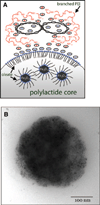
Figure 1
Figure 1
Magnetic nanoparticles (MNP) with surface complexed DNA. A) Schematically shown structure of a plasmid DNA-MNP complex based on polylactide plus oleate-coated iron oxide. MNP surface modification with polyethylenimine (PEI) via ion pairing with oleate enables formation of MNP-DNA complexes. For the sake of simplicity, only primary and a part of the secondary amines of branched PEI are shown protonated on the scheme. PEI complexaton of plasmid DNA results in formation of compact condensates (11). B) Transmission electron microscopy of MNP was performed after negative staining with 2% uranyl acetate (FEI Tecnai G2 electron microscope, Eindhoven, Netherlands). Note the small size and the large number of individual oleic acid-coated magnetite grains distributed in the MNP polymeric matrix.
Figure 2
Figure 2
Structural and physical characterization of MNP. A) MNP size as a function of THF amount in the organic phase. Insert shows the size distribution of the MNP. B) Zeta potential of DNA-complexing MNP as a function of the MNP/DNA weight ratio. Insert presents the effect of MNP/DNA weight ratio on DNA binding determined by PicoGreen assay. Varying MNP amounts were applied to 0.1 µg DNA and the unbound DNA was measured fluorimetrically (λexem=485 nm/530 nm). C) Magnetization curves of MNP formulations (125 µg samples). Note the absence of remanence (superparamagnetism) exhibited by the three types of MNP and the diamagnetic behavior of nonmagnetic nanoparticles measured as a control in an equivalent sample amount. Error bars indicate sd.
Figure 3
Figure 3
MNP uptake, GFP reporter expression, and cell viability in smooth muscle cells (A10) and bovine aortic endothelial cells (BAEC) (top and bottom rows, respectively) treated with three types of DNA complexing MNP, and nonmagnetic nanoparticles included as a control. MNP (1–5 µg per well) were incubated with DNA (0.25 µg/well) for 30 min and added to cells for 15 min under magnetic field. The uptake of MNP fluorescently labeled with PLA-BODIPY650/665 was determined fluorimetrically (λexem 620 nm/670 nm) in A10 cells and BAEC (A and B, respectively) after 24 h. Note the insignificant internalization of nonmagnetic nanoparticles compared with the MNP formulations. GFP expression of A10 (C) and BAEC (D) was determined fluorimetrically (485 nm/535 nm) in live cells 48 h post-treatment. Note the significantly higher transgene levels achieved with LMNP in both cell types compared with smaller sized MNP (P<0.01) and the marginal transfection mediated by nonmagnetic nanoparticles under the applied conditions (P≪0.001 vs. magnetic LMNP in both cell types). Viability of A10 (E) and BAEC (F) was determined 72 h post-treatment using the AlamarBlue assay. Error bars indicate sd.
Figure 4
Figure 4
MNP uptake (A, B), GFP reporter expression (C, D), and cell viability (E, F) in BAEC treated with DNA (0.25 µg/well) complexed with LMNP and applied to cells in low and high serum conditions (10% and 50% serum, respectively). (▲) Protocol 1: complexes applied under magnetic field for 15 min; (○) protocol 2: complexes applied without a magnetic field for 15 min; (□) protocol 3: complexes applied without a magnetic field for 2 h; (♦) protocol 4: complexes preincubated in the serum-containing medium for 2 h at 37°C and applied to cells under magnetic field for 15 min. Error bars indicate sd.
Figure 5
Figure 5
Internalization and trafficking of fluorescently labeled LMNP-DNA complexes examined by confocal microscopy in A10 (left panel) and BAEC (right panel). LMNP prepared using polylactide covalently labeled with BODIPY (FL) were incubated with Cy™ 5–labeled DNA (5 µg and 0.25 µg, respectively) for 30 min and applied to cells for 15 min under magnetic field. One hour before applying MNP-DNA complexes, cells were pretreated with Lysotracker Red at a concentration of 50 nM in DMEM supplemented with 10% FBS for lysosome staining. Cells images were obtained using three channels: green (λexem=488/492–519 nm), red (λexem=543/551–622 nm), and far red (λexem=633/640–754 nm) for MNP, lysosomes, and DNA, respectively. Note the cellular uptake of the complexes complete after 2 h, with little colocalization with the lysosomal compartment, and the substantial DNA dissociation from the carrier MNP in the cytosol 4 h after application.
Figure 6
Figure 6
Inhibitory effect of MNP-mediated adiponectin gene transfer on the growth factor-induced (PDGF-BB) proliferation of cultured smooth muscle cells (A10). Culture medium (DMEM) was supplemented with PDFG-BB at a concentration of 10 ng/ml and 2% of FBS. Growth inhibition of A10 seeded at 30% confluence mediated by adiponectin-encoding LMNP-DNA complexes (0.25 µg DNA/well) was measured in comparison to control complexes prepared with plasmids for GFP and GFP without a promoter.

References

    1. Barry ME, Pinto-Gonzalez D, Orson FM, McKenzie GJ, Petry GR, Barry MA. Role of endogenous endonucleases and tissue site in transfection and CpG-mediated immune activation after naked DNA injection. Hum. Gene Ther. 1999;10:2461–2480. - PubMed
    1. Lechardeur D, Sohn KJ, Haardt M, Joshi PB, Monck M, Graham RW, Beatty B, Squire J, O’Brodovich H, Lukacs GL. Metabolic instability of plasmid DNA in the cytosol: a potential barrier to gene transfer. Gene Ther. 1999;6:482–497. - PubMed
    1. Dean DA, Strong DD, Zimmer WE. Nuclear entry of nonviral vectors. Gene Ther. 2005;12:881–890. - PMC - PubMed
    1. Rolland A. Nuclear gene delivery: the Trojan horse approach. Expert Opin. Drug Deliv. 2006;3:1–10. - PubMed
    1. Scherer F, Anton M, Schillinger U, Henke J, Bergemann C, Krüger A, Gansbacher B, Plank C. Magnetofection: enhancing and targeting gene delivery by magnetic force in vitro and in vivo. Gene Ther. 2002;9:102–109. - PubMed

Publication types

LinkOut - more resources