Comparative Study
. 2007 Apr 10;104(15):6454-9.
doi: 10.1073/pnas.0610324104. Epub 2007 Apr 2.

Regulation of ryanodine receptor-dependent calcium signaling by polycystin-2

Affiliations
Comparative Study

Regulation of ryanodine receptor-dependent calcium signaling by polycystin-2

Georgia I Anyatonwu et al. Proc Natl Acad Sci U S A. .

Abstract

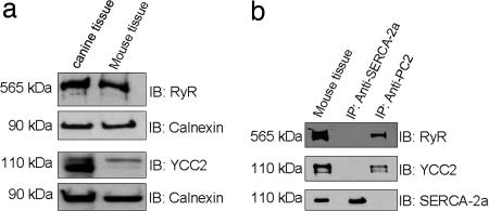
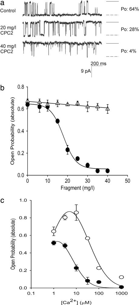
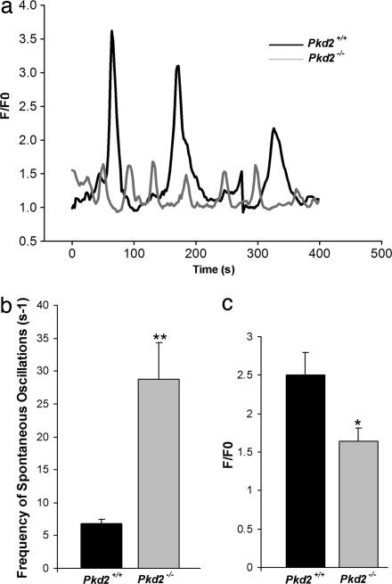
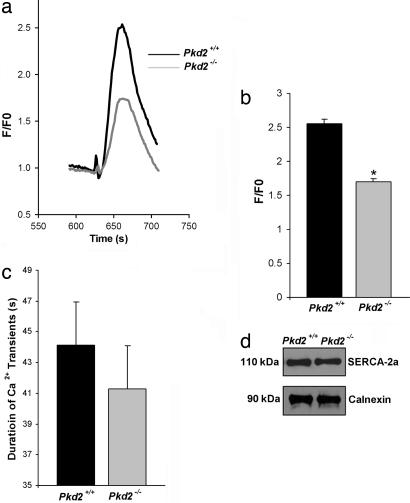
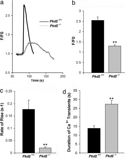
Mutations in polycystin-2 (PC2) cause autosomal dominant polycystic kidney disease. A function for PC2 in the heart has not been described. Here, we show that PC2 coimmunoprecipitates with the cardiac ryanodine receptor (RyR2) from mouse heart. Biochemical assays showed that the N terminus of PC2 binds the RyR2, whereas the C terminus only binds to RyR2 in its open state. Lipid bilayer electrophysiological experiments indicated that the C terminus of PC2 functionally inhibited RyR2 channel activity in the presence of calcium (Ca(2+)). Pkd2(-/-) cardiomyocytes had a higher frequency of spontaneous Ca(2+) oscillations, reduced Ca(2+) release from the sarcoplasmic reticulum stores, and reduced Ca(2+) content compared with Pkd2(+/+) cardiomyocytes. In the presence of caffeine, Pkd2(-/-) cardiomyocytes exhibited decreased peak fluorescence, a slower rate of rise, and a longer duration of Ca(2+) transients compared with Pkd2(+/+). These data suggest that PC2 is important for regulation of RyR2 function and that loss of this regulation of RyR2, as occurs when PC2 is mutated, results in altered Ca(2+) signaling in the heart.


Conflict of interest statement

The authors declare no conflict of interest.

Figures

Fig. 1.
Fig. 1.
Immunoprecipitation of RyR2 by PC2. (a) Expression of RyR2 and PC2 in canine and mouse heart. Lysates from canine and mouse heart were resolved on SDS/PAGE and immunoblotted (IB) with anti-RyR, anti-PC2 (YCC2), or anti-calnexin antibodies. Lysate, in lane 1, is from canine heart. Calnexin was used as the loading control in these experiments. The upper two panels are from the same blot, and the lower two panels are from the same blot. (b) Coimmunoprecipitation of RyR2 by PC2. Lysate isolated from mouse heart was subjected to immunoprecipitation (IP) with YCC2. Samples were blotted with antibodies to RyR2, PC2 (YCC2), and SERCA-2a. In these experiments, SERCA-2a was used as a negative control.
Fig. 2.
Fig. 2.
In vitro binding experiments. (a) Schematic representation of PC2 constructs used for in vitro binding experiments. (b) NPC2 binds the RyR2. In vitro binding experiments were performed with GST and HA fusion constructs as described in Materials and Methods. Samples were immunoblotted with antibodies to RyR2, GST, and HA. M indicates protein standard. (c) CPC2 binds to RyR2 in its open state. CPC2 was immobilized on glutathione beads and treated as described in Materials and Methods. Samples were immunoblotted (IB) with antibodies to RyR2 and GST. GST alone was used as for negative control. (d) N-terminal amino acid residues of PC2 that bind RyR2. A schematic representation of the N-terminal deletion constructs of PC2-L703X is shown. (e) PC2 N-terminal amino acid residues 130–220 bind the RyR2. Lysates from Madin-Darby canine kidney cells stably expressing mutants were resolved on SDS/PAGE, and the membrane containing proteins were immunoblotted by using anti-RyR and anti-HA antibodies.
Fig. 3.
Fig. 3.
The CPC2 inhibits RyR2 channel activity. (a) Cs+ currents obtained in the absence and presence of increasing concentration of GST-CPC2. Downward deflections represent channel openings. Experiments were performed under symmetrical conditions (250 mM CsCl in both the cis and trans side and 7–10 μM Ca2+ in the cis side). Currents traces were obtained at a holding potential of −25 mV and filtered at 400 Hz. To the right of each trace, the open state is represented as a dashed line, and the solid line represents the closed state. Open probability (Po) of channel at each concentration is shown on the right. (b) The CPC2 decreases the open probability of RyR2. Shown is concentration-dependent inhibition of RyR2 open probability when GST-CPC2 (filled circles; n = 3) and GST-NPC2 (open triangles; n = 3) is added to the cis side. Experiments were performed as described in Materials and Methods. (c) Ca2+-dependent inhibition of RyR2 by CPC2. The CPC2 decreases the open probability of RyR2 in the presence of increasing Ca2+ concentration (n = 3). Experiments were also performed in the absence (open circles) or presence (filled circles) of 17 μg/ml of GST-CPC2 (IC50) in the cis side before Ca2+ additions.
Fig. 4.
Fig. 4.
PC2 alters frequency and peak fluorescence of spontaneous oscillations in mouse cardiomyocytes. (a) Spontaneous oscillations of Pkd2+/+ and Pkd2−/− mouse cardiomyocytes. Representative traces of Ca2+ oscillations were monitored in the presence of Ca2+. The peak fluorescence for each genotype was measured, and each oscillation was normalized internally. The graph depicts the changes in fluorescence (F/F0) over time. (b) The frequency of spontaneous Ca2+ oscillations was determined by using a spectral analysis program in MATHLAB software (48). Statistical significance was calculated by using a one-tailed t test: ∗∗, P < 0.001 (n = 6 for Pkd2+/+; n = 12 for Pkd2−/−). (c) The change in peak fluorescence (F/F0) of each genotype was measured as described in Materials and Methods. Statistical significance was calculated by using a one-tailed t test: ∗, P < 0.05 (n = 28 for Pkd2+/+; n = 16 for Pkd2−/−).
Fig. 5.
Fig. 5.
PC2 required for maintenance the Ca2+ content in the SR stores. (a) Representative traces of Ca2+ transients in Pkd2+/+ and Pkd2−/− cardiomyocytes. Ca2+ transients were induced by application of 5 μM thapsigargin to the EGTA-buffered Ca2+-free extracellular solution. (b) Ca2+ content in the SR store. F/F0 was determined as described in Materials and Methods. Pkd2−/− mouse cardiomyocytes have a decreased Ca2+ content in the SR store. Statistical significance was calculated by using a one-tailed t test: ∗∗, P < 0.001 (n = 40 for Pkd2+/+; n = 40 for Pkd2−/−). (c) Duration of Ca2+ transient in Pkd2+/+ and Pkd2−/− cardiomyocytes. There was no difference in the duration of Ca2+ transients (n = 17 for Pkd2+/+ and n = 23 for Pkd2−/− cardiomyocytes). (d) SERCA-2a has similar expression levels in Pkd2+/+ and Pkd2−/− mouse cardiomyocytes. Tissue lysates were prepared from mouse heart at embryonic day 17.5 as described in Materials and Methods.
Fig. 6.
Fig. 6.
PC2 modulates the kinetics of Ca2+ transient from RyR2. (a) Representative traces of Ca2+ transient in Pkd2+/+ and Pkd2−/− cardiomyocytes. Cardiomyocytes were loaded with fluo-4, and Ca2+ transients were then elicited by 20 mM caffeine in the absence of extracellular Ca2+. (b) Comparison of the Ca2+ transient. Shown are F/F0 in Pkd2+/+ (n = 32) and Pkd2−/− (n = 40) cardiomyocytes. Statistical significance was calculated by using a one-tailed t test: ∗∗, P < 0.001. (c) Rate of rise of Ca2+ transient in Pkd2+/+ and Pkd2−/− cardiomyocytes. Pkd2−/− cardiomyocytes have significantly decreased peak in fluorescence compared with Pkd2+/+ cardiomyocytes: ∗∗, P = 0.001 (one-tailed t test). (d) Duration of Ca2+ transient in Pkd2+/+ and Pkd2−/− cardiomyocytes. Pkd2−/− cardiomyocytes had a significantly longer duration of Ca2+ transients compared with Pkd2+/+. Statistical significance was calculated by using a one-tailed t test: ∗∗, P < 0.001 (number of experiments for each genotype: 35).

References

    1. Gabow PA, Grantham JJ. In: Diseases of the Kidney. Schrier RW, Gottschalk CW, editors. Boston: Little, Brown; 1997. pp. 535–569.
    1. Belz MM, Fick-Brosnahan GM, Hughes RL, Rubinstein D, Chapman AB, Johnson AM, McFann KK, Kaehny WD, Gabow PA. Kidney Int. 2003;63:1824–1830. - PubMed
    1. Chapman AB, Johnson AM, Rainguet S, Hossack K, Gabow P, Schrier RW. J Am Soc Nephrol. 1997;8:1292–1297. - PubMed
    1. Igarashi P, Somlo S. J Am Soc Nephrol. 2002;13:2384–2398. - PubMed
    1. Calvet JP. J Nephrol. 1998;11:24–34. - PubMed

Publication types