Comparative Study
. 2007 Apr 10;104(15):6400-5.
doi: 10.1073/pnas.0701579104. Epub 2007 Apr 2.

Lipoprotein SmpA is a component of the YaeT complex that assembles outer membrane proteins in Escherichia coli

Affiliations
Comparative Study

Lipoprotein SmpA is a component of the YaeT complex that assembles outer membrane proteins in Escherichia coli

Joseph G Sklar et al. Proc Natl Acad Sci U S A. .

Abstract

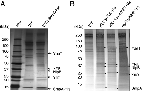
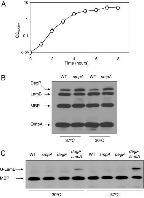
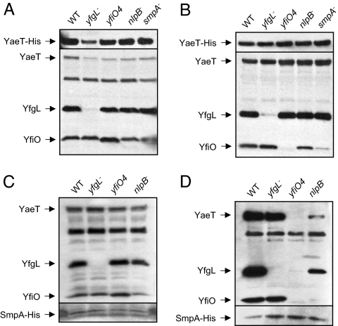
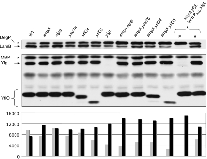
A major role of the outer membrane (OM) of Gram-negative bacteria is to provide a protective permeability barrier for the cell, and proper maintenance of the OM is required for cellular viability. OM biogenesis requires the coordinated assembly of constituent lipids and proteins via dedicated OM assembly machineries. We have previously shown that, in Escherichia coli, the multicomponent YaeT complex is responsible for the assembly of OM beta-barrel proteins (OMPs). This complex contains the OMP YaeT and three OM lipoproteins. Here, we report another component of the YaeT complex, the OM lipoprotein small protein A (SmpA). Strains carrying loss-of-function mutations in smpA are viable but exhibit defects in OMP assembly. Biochemical experiments show that SmpA is involved in maintaining complex stability. Taken together, these experiments establish an important role for SmpA in both the structure and function of the YaeT complex.


Conflict of interest statement

The authors declare no conflict of interest.

Figures

Fig. 1.
Fig. 1.
SmpA copurifies with the YaeT complex. (A) Silver-stained SDS/polyacrylamide gel of samples from cell lysates of wild-type (WT), wt/pSmpA-His strains immunoprecipitated with anti-His tag antibody. (B) Silver-stained SDS/polyacrylamide gel of samples from cell lysates of WT, yfgL/pYfgL-His, yfiO::kan/pYfiO-His, and nlpB/pNlpB-His strains immunoprecipitated with anti-His tag antibody. MW, molecular weight.
Fig. 2.
Fig. 2.
SmpA is not essential for growth in E. coli. (A) Growth curves for wild-type cells (♦) or smpA cells (▫) at 37°C are shown. Growth was monitored by measuring the optical density at 600 nm every hour for 8 h. The results of a representative experiment are shown above. (B) Levels of envelope proteins in an smpA mutant during midlogarithmic growth at 37°C and 30°C. Western blot analysis shows a very small reduction in the levels of OmpA and LamB in an smpA mutant strain when compared with a wild-type (WT) strain. Levels of the periplasmic chaperone/protease DegP are elevated 1.5- to 2-fold in an smpA mutant. (C) Unfolded LamB accumulates in an smpA degP double mutant. Cells were grown to midlogarithmic growth at 37°C and 30°C and harvested by centrifugation. These samples were gently lysed to preserve the native conformation of OMPs. Western blot analysis by using LamB antiserum was used to visualize unfolded LamB (U-LamB). Levels of MBP are shown as a loading control.
Fig. 3.
Fig. 3.
Affinity purification of the YaeT complex in lipoprotein mutants. (A–D) Affinity purification was performed in WT and lipoprotein mutant strains that contain pYaeT-His (A and B) and pSmpA-His (C and D), respectively. (A and C) Protein levels in the total membrane fraction isolated from cells before any purification. (B and D) Samples after they have passed through an Ni-affinity column. Samples were blotted against antibodies that recognize YaeT, YfgL, and YfiO and the His tag.
Fig. 4.
Fig. 4.
smpA genetic interactions. Western blot analysis shows the levels of DegP, LamB, MBP, YfgL, and YfiO proteins in strains carrying mutations that affect different YaeT complex members and in smpA double mutants during midlogarithmic growth at 30°C. The smpA yfgL depletion strain was grown in the presence of l-arabinose (A) to induce expression of yfgL or in the presence of d-fucose (F) to prevent its expression. Below the Western blots is a chart that shows the quantification of LamB (gray bars) and DegP (black bars). The values of the y axis represent the areas of the bands as quantified by using ImageJ image-processing software.

References

    1. Kamio Y, Nikaido H. Biochemistry. 1976;15:2561–2570. - PubMed
    1. Ruiz N, Kahne D, Silhavy TJ. Nat Rev Microbiol. 2006;4:57–66. - PubMed
    1. Narita S, Tokuda H. FEBS Lett. 2006;580:1164–1170. - PubMed
    1. Wu T, Malinverni J, Ruiz N, Kim S, Silhavy TJ, Kahne D. Cell. 2005;121:235–245. - PubMed
    1. Bos MP, Tefsen B, Geurtsen J, Tommassen J. Proc Natl Acad Sci USA. 2004;101:9417–9422. - PMC - PubMed

Publication types

MeSH terms

LinkOut - more resources