Comparative Study
. 2007 Apr 10;104(15):6247-52.
doi: 10.1073/pnas.0701509104. Epub 2007 Apr 2.

Wnt signaling regulates pancreatic beta cell proliferation

Affiliations
Comparative Study

Wnt signaling regulates pancreatic beta cell proliferation

Ingrid C Rulifson et al. Proc Natl Acad Sci U S A. .

Abstract

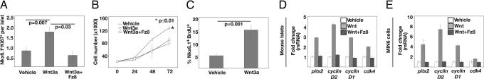
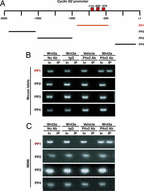
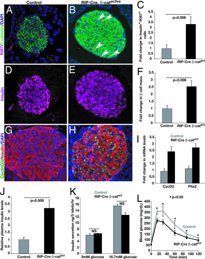
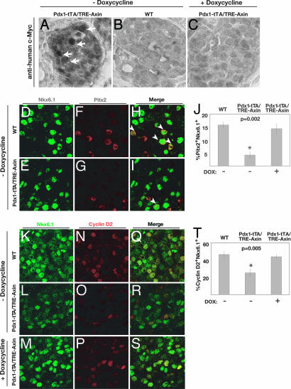
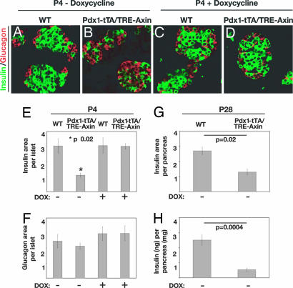
There is widespread interest in defining factors and mechanisms that stimulate proliferation of pancreatic islet cells. Wnt signaling is an important regulator of organ growth and cell fates, and genes encoding Wnt-signaling factors are expressed in the pancreas. However, it is unclear whether Wnt signaling regulates pancreatic islet proliferation and differentiation. Here we provide evidence that Wnt signaling stimulates islet beta cell proliferation. The addition of purified Wnt3a protein to cultured beta cells or islets promoted expression of Pitx2, a direct target of Wnt signaling, and Cyclin D2, an essential regulator of beta cell cycle progression, and led to increased beta cell proliferation in vitro. Conditional pancreatic beta cell expression of activated beta-catenin, a crucial Wnt signal transduction protein, produced similar phenotypes in vivo, leading to beta cell expansion, increased insulin production and serum levels, and enhanced glucose handling. Conditional beta cell expression of Axin, a potent negative regulator of Wnt signaling, led to reduced Pitx2 and Cyclin D2 expression by beta cells, resulting in reduced neonatal beta cell expansion and mass and impaired glucose tolerance. Thus, Wnt signaling is both necessary and sufficient for islet beta cell proliferation, and our study provides previously unrecognized evidence of a mechanism governing endocrine pancreas growth and function.


Conflict of interest statement

The authors declare no conflict of interest.

Figures

Fig. 1.
Fig. 1.
Purified Wnt3a induces expression of Pitx2, cyclin D2, and other cell cycle regulators in β cells and promotes cell expansion. (A) Expression of Ki67 in Nkx6.1+ cells of purified WT P8 islets stimulated with Wnt3a, vehicle, or Wnt3a + Fz8-CRD. (B) Growth of MIN6 cells stimulated with vehicle only, Wnt3a, or Wnt3a + Fz8-CRD and counted at 24, 48, and 72 h. Each condition was performed in triplicate. (C) MIN6 cells stimulated with Wnt3a or vehicle for 8 h were assayed for BrdU incorporation by immunofluorescence analysis. (A–C) Data are presented as the average ± SEM. All RT-PCR results were normalized to β-actin and are the average value from triplicate experiments. P values are indicated in each graph. (D) Real-time RT-PCR analysis of Pitx2, cyclin D2, cyclin D1, and cdk4 cDNA from purified P8 islets treated with vehicle only, purified Wnt3a, or Wnt3a + Fz8-CRD for 24 h. (E) Real-time RT-PCR analysis of Pitx2, cyclin D2, cyclin D1, and cdk4 cDNA from MIN6 cells treated with vehicle only, purified Wnt3a, or Wnt3a + Fz8-CRD for 24 h.
Fig. 2.
Fig. 2.
ChIP demonstrates that Pitx2 associates with specific elements in the cyclin D2 promoter upon Wnt3a stimulation. (A) Position of DNA regions flanked by primer pairs (PP) used for PCR to assess Pitx2 association with elements within the cyclin D2 promoter. Consensus bicoid binding sequences are marked by red boxes. (B) Anti-Pitx2 ChIP performed on 8-h Wnt3a-stimulated islet cultures derived from WT P8 mice reveals Pitx2 binding to the cyclin D2 promoter at sites −980 to −525 upstream of the transcriptional start site. (C) Anti-Pitx2 ChIP performed on MIN6 cells treated with Wnt3a for 3 h and subsequently analyzed with PCR shows a similar binding pattern to the cyclin D2 promoter.
Fig. 3.
Fig. 3.
Conditional expression of activated β-catenin in RIP-Cre, β-catactive islets. (A and B) Elevated β-catenin expression and an increased number of proliferating Ki67+ cells in RIP-Cre, β-catactive islets compared with controls. (C) Quantification of insulin+, Ki67+ cells in control and RIP-Cre, β-catactive islets. (D and E) Immunostaining reveals insulin expression is maintained in proliferating Ki67+ cells in RIP-Cre, β-catactive islets. (F) Quantification of pancreatic β cell mass in control and RIP-Cre, β-catactive mice. (G and H) Immunostaining reveals increased numbers of cyclin D2-expressing insulin+ cells in RIP-Cre, β-catactive islets. (I) Real-time PCR quantification of cyclin D2 mRNA levels in isolated control and RIP-Cre, β-catactive islets. All data are statistically significant. P < 0.005. (J) Serum insulin levels in fed control and RIP-Cre, β-catactive mice. (K) Assessment of insulin secretion in response to increased glucose as performed on cultured islets. P values were not significant (NS). (L) i.p. glucose tolerance tests of control and RIP-Cre, β-catactive mice after overnight fasting. (J–L) Values represent mean ± SEM. P values are indicated where appropriate.
Fig. 4.
Fig. 4.
Dox-dependent expression of Axin, Pitx2, and Cyclin D2 in Pdx1-tTA/TRE-Axin islets. (A–C) Immunohistochemical detection of Myc-tagged Axin expression in islets. Pdx1-tTA/TRE-Axin P4 mice (A) express Myc-tagged Axin (arrows), which is absent in WT (B) and Pdx1-tTA/TRE-Axin (+ Doxycycline) (C) control mice at P4. (Original magnification: ×63.) (D–I) Immunofluorescent detection of Nkx6.1 (green), Pitx2 (red), and merge (yellow) in the islets of P4 WT and Pdx1-tTA/TRE-Axin mice and Pdx1-tTA/TRE-Axin mice on Dox from the time of conception. (H and I) White arrowheads indicate Nkx6.1+ /Pitx2+ nuclei. (J) Percentage of Nkx6.1+ that are Pitx2+ in Pdx1-tTA/TRE-Axin and control mice. (K–S) Immunofluorescent detection of Nkx6.1 (green) and Cyclin D2 (red) in the islets of P4 WT and Pdx1-tTA/TRE-Axin mice and Pdx1-tTA/TRE-Axin mice on Dox. (T) Percentage of Nkx6.1+ that are Cyclin D2+ in Pdx1-tTA/TRE-Axin and control mice. Data are presented as the average ± SEM. P values are indicated in the figures. Data are from at least five mice per genotype. (Original magnification: ×100.)
Fig. 5.
Fig. 5.
Conditional Axin expression in Pdx1+ progenitors impairs β cell development. (A–D) Immunofluorescent detection of insulin+ (green) and glucagon+ (red) cells in WT (A) and Pdx1-tTA/TRE-Axin (B) islets (no Dox) and in WT (C) and Pdx1-tTA/TRE-Axin (D) islets administered Dox from the time of conception. (Original magnification: ×100.) (E and F) The relative insulin+ (E) and glucagon (F) area per islet compared in WT and Pdx1-tTA/TRE-Axin mice and in WT and Pdx1-tTA/TRE-Axin mice administered Dox from the time of conception. (E) The P value for insulin+ area between Pdx1-tTA/TRE-Axin islets and control islets is ≤0.02. (F) The P value for glucagon+ area among all genotypes is not significant. (G) Morphometric analysis of insulin+ area as a percentage of total pancreas area from P28 WT and Pdx1-tTA/TRE-Axin mice. (H) Total insulin (nanograms) per pancreas (milligrams) in P28 Pdx1-tTA/TRE-Axin and WT mice. All data are from at least three litters, with four to eight mice per genotype. Data are presented as the average ± SEM.

References

    1. Butler AE, Janson J, Bonner-Weir S, Ritzel R, Rizza RA, Butler PC. Diabetes. 2003;52:102–110. - PubMed
    1. Ryan EA, Paty BW, Senior PA, Bigam D, Alfadhli E, Kneteman NM, Lakey JR, Shapiro AM. Diabetes. 2005;54:2060–2069. - PubMed
    1. Heit JJ, Karnik SK, Kim SK. Annu Rev Cell Dev Biol. 2006;22:311–338. - PubMed
    1. Cozar-Castellano I, Fiaschi-Taesch N, Bigatel TA, Takane KK, Garcia-Ocana A, Vasavada R, Stewart AF. Endocr Rev. 2006;27:356–370. - PubMed
    1. Logan CY, Nusse R. Annu Rev Cell Dev Biol. 2004;20:781–810. - PubMed

Publication types

LinkOut - more resources