

Migration of intradermally injected quantum dots to sentinel organs in mice
- PMID: 17404394
- PMCID: PMC3471152
- DOI: 10.1093/toxsci/kfm074
Migration of intradermally injected quantum dots to sentinel organs in mice
Abstract
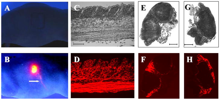
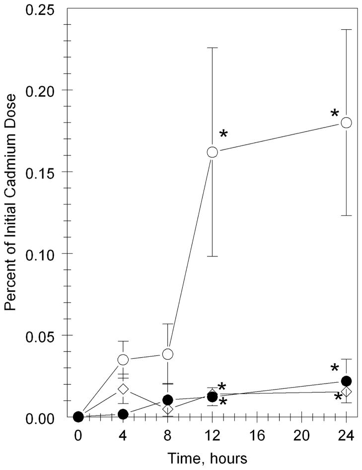
Topical exposure to nanoscale materials is likely from a variety of sources including sunscreens and cosmetics. Because the in vivo disposition of nanoscale materials is not well understood, we have evaluated the distribution of quantum dots (QDs) following intradermal injection into female SKH-1 hairless mice as a model system for determining tissue localization following intradermal infiltration. The QD (CdSe core, CdS capped, poly[ethylene glycol] coated, 37 nm diameter, 621 nm fluorescence emission) were injected intradermally (ID) on the right dorsal flank. Within minutes following intradermal injection, the highly UV fluorescent QD could be observed moving from the injection sites apparently through the lymphatic duct system to regional lymph nodes. Residual fluorescent QD remained at the site of injection until necropsy at 24 h. Quantification of cadmium and selenium levels after 0, 4, 8, 12, or 24 h in multiple tissues, using inductively coupled plasma mass spectrometry (ICP-MS), showed a time-dependent loss of cadmium from the injection site, and accumulation in the liver, regional draining lymph nodes, kidney, spleen, and hepatic lymph node. Fluorescence microscopy corroborated the ICP-MS results regarding the tissue distribution of QD. The results indicated that (1) ID injected nanoscale QD remained as a deposit in skin and penetrated the surrounding viable subcutis, (2) QD were distributed to draining lymph nodes through the sc lymphatics and to the liver and other organs, and (3) sentinel organs are effective locations for monitoring transdermal penetration of nanoscale materials into animals.
Figures


References
-
- Aleboyeh A, Aleboyeh H, Moussa Y. “Critical” effect of hydrogen peroxide in photochemical oxidative decolorization of dyes: Acid Orange 8, Acid Blue 74 and Methyl Orange. Dyes and Pigments. 2003;57:67–75.
-
- Alivisatos AP, Gu W, Larabell C. Quantum dots as cellular probes. Annu Rev Biomed Eng. 2005;7:55–76. - PubMed
-
- Al-Somali AM, Kreuger KM, Falkner JC, Colvin VL. Recycling size exclusion chromatography for the analysis and separation of nanocrystalline gold. Analy Chem. 2004;76:5903–5910. - PubMed
-
- Azzazy HME, Mansour MMH, Kazmierczak SC. Nanodiagnostics: A new frontier in clinical laboratory medicine. Clin Chem. 2006;52:1238–1246. - PubMed
Publication types
MeSH terms
Substances
Grants and funding
LinkOut - more resources
Full Text Sources
Other Literature Sources
