. 2007 Apr;117(4):1068-77.
doi: 10.1172/JCI30117.

Hypoxia-inducible factor-2 (HIF-2) regulates hepatic erythropoietin in vivo

Affiliations

Hypoxia-inducible factor-2 (HIF-2) regulates hepatic erythropoietin in vivo

Erinn B Rankin et al. J Clin Invest. 2007 Apr.

Abstract

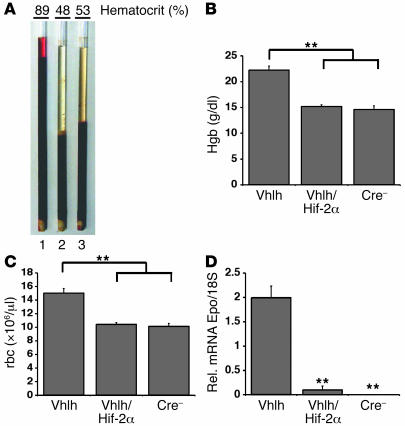
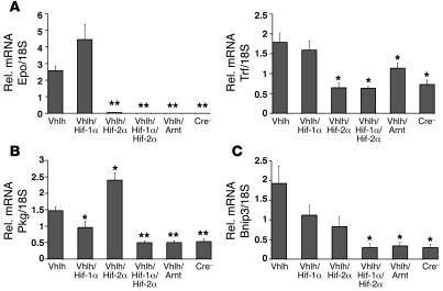
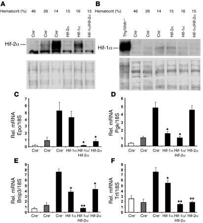
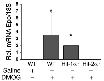
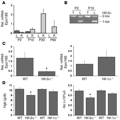
Erythropoiesis is critically dependent on erythropoietin (EPO), a glycoprotein hormone that is regulated by hypoxia-inducible factor (HIF). Hepatocytes are the primary source of extrarenal EPO in the adult and express HIF-1 and HIF-2, whose roles in the hypoxic induction of EPO remain controversial. In order to define the role of HIF-1 and HIF-2 in the regulation of hepatic EPO expression, we have generated mice with conditional inactivation of Hif-1alpha and/or Hif-2alpha (Epas1) in hepatocytes. We have previously shown that inactivation of the von Hippel-Lindau tumor suppressor pVHL, which targets both HIFs for proteasomal degradation, results in increased hepatic Epo production and polycythemia independent of Hif-1alpha. Here we show that conditional inactivation of Hif-2alpha in pVHL-deficient mice suppressed hepatic Epo and the development of polycythemia. Furthermore, we found that physiological Epo expression in infant livers required Hif-2alpha but not Hif-1alpha and that the hypoxic induction of liver Epo in anemic adults was Hif-2alpha dependent. Since other Hif target genes such phosphoglycerate kinase 1 (Pgk) were Hif-1alpha dependent, we provide genetic evidence that HIF-1 and HIF-2 have distinct roles in the regulation of hypoxia-inducible genes and that EPO is preferentially regulated by HIF-2 in the liver.


Figures

Figure 1
Figure 1. Generation of mice deficient for Vhlh, Vhlh/Hif-1α, Vhlh/Hif-2α, and Vhlh/ Hif-1α/Hif-2α in the liver.
(A) Genomic maps of the Vhlh (2-lox), Hif-1α (2-lox), and Hif-2α (2-lox) conditional alleles. Numbered boxes represent exons targeted for deletion. The locations of LoxP sites and primers used to amplify the conditional allele (2-lox), recombined allele (1-lox), and WT allele are shown with gray triangles and arrows, respectively. N, NcoI; E, EcoR1; P, PstI. (B) Genomic PCR analysis of DNA isolated from the tail (T) and liver (L) of PEPCK-Cre mutant mice. (C) Genomic PCR analysis of DNA isolated from the tail and liver of 4-week-old albumin-Cre mutant mice. Note that mice were bred in a mixed genetic background. Occasionally 2 different Hif-2α WT alleles were detected by genomic PCR analysis, suggesting a polymorphism in the amplified region. (D) Western blot analysis of nuclear protein isolated from livers of the following albumin-Cre mutant littermates: lane 1, -Vhlh haploinsufficient for Hif-1α; lane 2, -Vhlh/Hif-1α haploinsufficient for Hif-2α; lane 3, -Vhlh/Hif-2α; lane 4, -Vhlh/Hif-1α/Hif-2α; lane 5, -Vhlh/Arnt; and lane 6, Cre-negative mice. Note that the recombination efficiency of the Hif-1α conditional allele is best determined by comparing Cre-negative control mice to Vhlh/Hif-1α/Hif-2α mutant mice due to the presence of nonrecombined inflammatory cells in albumin-Vhlh/Hif-1α mutant mice.
Figure 2
Figure 2. Inactivation of Hif-2α suppresses the development of polycythemia in PEPCK-Vhlh mutant mice.
(A) Photograph of centrifuged microcapillary tubes containing blood from representative PEPCK-Vhlh (hematocrit 89%; 1), PEPCK-Vhlh/Hif-2α (hematocrit 48%; 2), and control (hematocrit 53%; 3) mice. (B and C) Elevated hemoglobin (Hgb) and red blood cell numbers in PEPCK-Vhlh mutant mice were suppressed in PEPCK-Vhlh/Hif-2α mutant mice. Shown are hemoglobin concentrations and red blood cell (rbc) numbers in blood collected from PEPCK-Cre mutant and control (Cre) mice determined by a complete blood count analyzer. Shown are the mean values for 6 individual mice. Error bars represent SEM. (D) Inactivation of Hif-2α significantly decreases Epo transcript levels in PEPCK-Vhlh mutant livers. Shown are relative Epo mRNA transcript levels normalized to 18S in the livers of PEPCK-Cre mutant and Cre-negative mice. Bars represent mean mRNA transcript levels of 3 mice per group. Error bars represent SEM. **P < 0.001 compared with PEPCK-Vhlh as determined by Student’s t test.
Figure 3
Figure 3. Differential HIF target gene expression in albumin-Vhlh/Hif-1α– and albumin-Vhlh/Hif-2α–deficient livers.
Relative mRNA levels for HIF target genes in the liver of albumin-Cre mutant mice determined by real-time PCR. (A) Inactivation of Hif-2α significantly decreases the expression of HIF target genes regulating erythropoiesis (Epo) and iron transport (Trf). (B) Inactivation of Hif-1α decreases the expression of the glycolytic target gene Pgk. (C) Inactivation of both Hif-1α and Hif-2α decreases the expression of the proapoptotic gene Bnip3. Bars represent the mean mRNA transcript level of 3 mice for the albumin-Vhlh/Hif-1α/Hif-2α group and 4 mice for all other groups. Error bars represent SEM. *P < 0.05, **P < 0.001 compared with albumin-Vhlh mutants as determined by Student’s t test.
Figure 4
Figure 4. Hepatic Epo expression in anemic mice is Hif-2α dependent.
(A and B) Hif-1α and Hif-2α proteins are stabilized in the livers of anemic mice as determined by Western blot analysis. Nuclear protein extracts were isolated from livers of albumin-Cre mutant mice of the indicated genotypes. Nuclear protein extracts isolated from the thymus (Thy) of Vhlh-deficient mice was used as a positive Hif-1α protein control. Ponceau S staining is shown to demonstrate equal protein loading. (CF) Real-time PCR analysis of hepatic Epo, Pgk, Bnip3, and Trf expression in anemic albumin-Cre mutant mice. White bars represent mice with hematocrit values of approximately 45%; gray bars, approximately 27%; and black bars, approximately 15%. Bars represent average mRNA transcript levels of 3 mice for each group; error bars indicate SEM. *P < 0.05, **P < 0.001 compared with control mice (Cre) with the same range of hematocrit as determined by Student’s t test.
Figure 5
Figure 5. Inhibition of Hif prolyl hydroxylation results in an Hif-2–dependent induction of hepatic Epo.
Relative Epo mRNA levels in the liver of albumin-Cre mutant mice treated with either with normal saline or 10 mg of DMOG as determined by real-time PCR. Bars represent the mean Epo mRNA transcript level of 3 mice for the WT and albumin–Hif-1α mutants (Hif-1α–/–) and 5 mice for the albumin-Hif-2α (Hif-2α–/–) mutants. Error bars represent SEM. *P < 0.05 compared with albumin–Hif-2α mutant mice as determined by the Wilcoxon rank sum test.
Figure 6
Figure 6. Hif-2 regulates hepatic Epo expression in infant mice.
(A) Relative Epo mRNA levels in the livers and kidneys (K) of P2, P10, P20, and P60 mice. Bars represent the average Epo mRNA transcript level of 3 mice for each condition. (B) Genomic PCR analysis of DNA isolated from the tail and liver of albumin–Hif-2α mutant mice at P2 and P10. Note that the ratio of the recombined (1-lox) allele to the unrecombined (2-lox) Hif-2α allele increased between P2 and P10. (C) Relative Epo mRNA expression levels in P10 albumin–Hif-2α (Hif-2α–/–) and albumin–Hif-1α (Hif-1α–/–) and Cre-recombinase–negative control littermates (WT). Bars represent the mean Epo mRNA transcript level for WT albumin–Hif-2α littermates (n = 12) and albumin–Hif-2α mutants (n = 13) (left) and albumin–Hif-1α mutants and littermate controls (n = 10 each) (left). (D) Hemoglobin concentrations and red blood cell numbers in blood collected from 10-day-old albumin-Cre mutant and control mice. Bars represent the mean values for albumin–Hif-2α controls and mutants (n = 12 each), albumin–Hif-1α controls (n = 18), and albumin–Hif-1α mutants (n = 11). Error bars represent SEM. *P < 0.05 compared with littermate controls as determined by Student’s t test.
Figure 7
Figure 7. HIF-2α preferentially binds to the endogenous EPO 3′ HRE in hepatocytes.
(A) Western blot analysis for HIF-1 and HIF-2 in normoxic (21% O2; NX) and hypoxic (1% O2; HX) Hep3B cells. (B) EPO and PGK1 mRNA levels in normoxic and hypoxic Hep3B cells as determined by real-time PCR. (C) The hypoxic induction of EPO expression in Hep3B cells is HIF-2 dependent. Real-time PCR analysis of EPO expression in normoxic and hypoxic Hep3B cells treated with control, HIF-1α, HIF-2α, or control siRNA oligonucleotides. Error bars represent SD. (D) HIF-1α preferentially binds to the EPO HRE in vitro as determined by EMSA. s.s.HIF, HIF supershift. + and – indicate the presence and absence, respectively, of the antibody used in a supershift reaction. (E) ChIP analysis of the EPO and PGK1 HREs in normoxic and hypoxic Hep3B cells using antibodies directed against HIF-1α, HIF-2α, and CBP/p300. Coprecipitated DNA fragments were detected by PCR using primers spanning the EPO and PGK1 HREs. mAb-HIF-1α, mAb against HIF-1α; pAb-HIF-2α, polyclonal antibody against human HIF-2α; pAb-p300, polyclonal antibody against human p300.

Comment in

References

    1. Ebert B.L., Bunn H.F. Regulation of the erythropoietin gene. Blood. 1999;94:1864–1877. - PubMed
    1. Wu H., Lee S.H., Gao J., Liu X., Iruela-Arispe M.L. Inactivation of erythropoietin leads to defects in cardiac morphogenesis. Development. 1999;126:3597–3605. - PubMed
    1. Wu H., Liu X., Jaenisch R., Lodish H.F. Generation of committed erythroid BFU-E and CFU-E progenitors does not require erythropoietin or the erythropoietin receptor. Cell. 1995;83:59–67. - PubMed
    1. Zanjani E.D., Ascensao J.L., McGlave P.B., Banisadre M., Ash R.C. Studies on the liver to kidney switch of erythropoietin production. J. Clin. Invest. 1981;67:1183–1188. - PMC - PubMed
    1. Dame C., et al. Erythropoietin mRNA expression in human fetal and neonatal tissue. Blood. 1998;92:3218–3225. - PubMed

Publication types

MeSH terms

Substances