. 2007 Apr;117(4):1088-95.
doi: 10.1172/JCI31242.

Local production of angiotensin II in the subfornical organ causes elevated drinking

Affiliations

Local production of angiotensin II in the subfornical organ causes elevated drinking

Koji Sakai et al. J Clin Invest. 2007 Apr.

Abstract

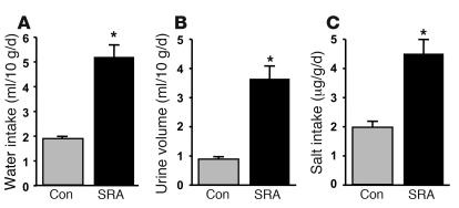
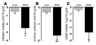
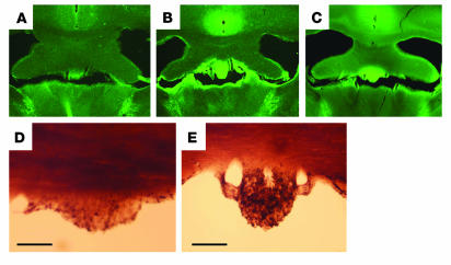
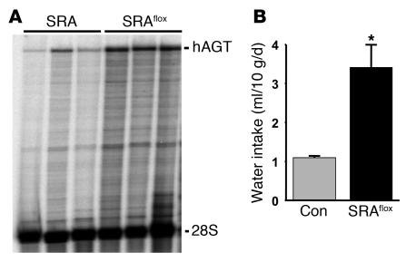
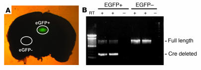
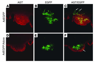
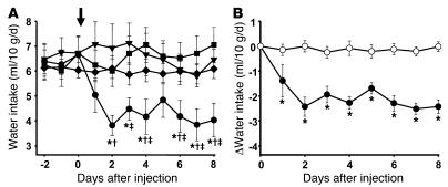
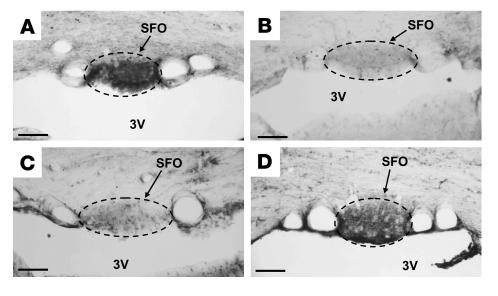
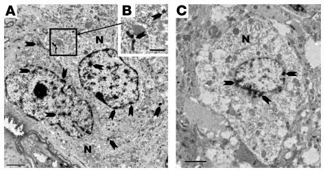
The mechanism controlling cell-specific Ang II production in the brain remains unclear despite evidence supporting neuron-specific renin and glial- and neuronal-specific angiotensinogen (AGT) expression. We generated double-transgenic mice expressing human renin (hREN) from a neuron-specific promoter and human AGT (hAGT) from its own promoter (SRA mice) to emulate this expression. SRA mice exhibited an increase in water and salt intake and urinary volume, which were significantly reduced after chronic intracerebroventricular delivery of losartan. Ang II-like immunoreactivity was markedly increased in the subfornical organ (SFO). To further evaluate the physiological importance of de novo Ang II production specifically in the SFO, we utilized a transgenic mouse model expressing a floxed version of hAGT (hAGT(flox)), so that deletions could be induced with Cre recombinase. We targeted SFO-specific ablation of hAGT(flox) by microinjection of an adenovirus encoding Cre recombinase (AdCre). SRA(flox) mice exhibited a marked increase in drinking at baseline and a significant decrease in water intake after administration of AdCre/adenovirus encoding enhanced GFP (AdCre/AdEGFP), but not after administration of AdEGFP alone. This decrease only occurred when Cre recombinase correctly targeted the SFO and correlated with a loss of hAGT and angiotensin peptide immunostaining in the SFO. These data provide strong genetic evidence implicating de novo synthesis of Ang II in the SFO as an integral player in fluid homeostasis.


Figures

Figure 1
Figure 1. Volume homeostasis in SRA mice.
Water intake (A), urine volume (B), and salt intake (C) in SRA mice (n = 14) and control littermates (Con; n = 25). *P < 0.001 compared with control mice.
Figure 2
Figure 2. The effect of chronic icv administration of losartan on volume homeostasis in SRA mice.
Change in water intake (A), urine volume (B), and salt intake (C) in SRA mice (n = 9) and nontransgenic mice (n = 6) in response to losartan administered icv. *P = 0.007 (A); **P < 0.001 (B); #P = 0.002 (C) compared with control mice.
Figure 3
Figure 3. Localization of angiotensin peptide in the brain of SRA and SRAflox mice.
Representative photomicrographs of immunostaining for Ang I/II peptide in the SFO by immunofluorescence (AC) and immunoperoxidase (D and E) in nontransgenic control (A and D), SRA (B and E), and SRAflox (C) mice. n = 4 or more mice. Scale bars: 0.4 mm (AC); 0.1 mm (D and E).
Figure 4
Figure 4. Expression hAGT in the brain and water intake in SRAflox mice.
(A) An RNase protection assay of total RNA purified from independent brain samples removed from 3 SRA and 3 SRAflox mice. The positions of the hAGT- and 28S-protected bands are shown. (B) Water intake in SRAflox (n = 4) and age- and sex-matched littermate controls (n = 9). *P < 0.001 compared with control mice.
Figure 5
Figure 5. Site-specific deletion of hAGT mRNA in the SFO.
(A) Representative brain slice from an SRAflox mouse coinfected with AdEGFP and AdCre and photographed under a fluorescence stereomicroscope. Selected from over 12 photos each from independent mice (n = 12). Brain tissue from an EGFP-positive region and from a control region (EGFP–) were carefully dissected for the experiment in B. (B) RT-PCR reaction for hAGT mRNA from brain punches derived from the EGFP-positive and -negative regions indicated in A.
Figure 6
Figure 6. Site-specific deletion of hAGT protein in the SFO.
Representative coronal sections through the SFO from mice injected with either AdEGFP alone or both AdEGFP and AdCre directly into the SFO. AGT was localized by immunofluorescence (red) and EGFP by its intrinsic fluorescence (green). AC show colocalization of AdEGFP and AGT but no deletion or diminution of AGT staining. The arrows in C indicate EGFP-positive, hAGT-negative cells adjacent to the SFO. DF show loss of AGT in regions where the virus mixture (AdEGFP and AdCre) is located. The arrow in F represents a cell in which hAGT was not ablated, which may represent a cell infected with AdEGFP but not AdCre.
Figure 7
Figure 7. Effect of AdCre transfection into the SFO on water intake.
(A) Drinking volume of water before and after SFO-directed injection (arrow) of AdCre/AdEGFP into SRA mice lacking loxP sites (filled squares; n = 8); AdEGFP alone into SRAflox mice (filled triangles; n = 8); AdCre/AdEGFP into SRAflox mice, where the injection missed the SFO (filled diamonds; n = 21); and AdCre/AdEGFP into SRAflox mice, where the injection into the SFO was confirmed (filled circles; n = 8). Data were analyzed using 2-way repeated measures ANOVA with the Bonferroni posthoc test. *P < 0.05 versus SRAflox mice injected with AdEGFP; P < 0.05 versus SRAflox mice injected with AdCre, which missed the SFO; P < 0.05 versus SRA mice lacking loxP sites injected with AdCre/AdEGFP. (B) Change in water intake by mice after SFO-directed injection of AdCre/AdEGFP into SRAflox mice, where the injection missed the SFO (open circles) and of AdCre/AdEGFP into SRAflox mice, where the injection into the SFO was confirmed (filled circles). *P < 0.003.
Figure 8
Figure 8. Effect of AdCre on SFO Ang II.
Immunocytochemistry for angiotensin I/II in the SFO of SRAflox mice. (A) SRAflox mouse injected with AdEGFP alone. (B) Diminished Ang I/II signal in SFO after SFO-directed injection of AdCre/AdEGFP. (C) Control SFO section from SRAflox mice where the primary antibody directed against Ang I/II was omitted. (D) Retention of Ang I/II immunoreactivity in SFO after an injection of AdCre/AdEGFP that missed the SFO. The SFO is bordered by a dashed line. 3V, third ventricle. Scale bars: 100 μm.
Figure 9
Figure 9. Ultrastructural localization of Ang I/II in the SFO.
(A) Strong expression of Ang I/II (arrows) in the nucleus, cytosol, and vesicular structures of SFO neurons of an uninjected SRAflox mouse. (B) Fragment of cytoplasm of an SFO neuron in A containing immunoreactive vesicular structures (arrows). (C) Note the very low level of immunoreactivity around the neuronal nucleus (arrows) and absence of immunoreactivity in cytoplasm and neuropil after AdCre/AdEGFP was directly injected into the SFO. N, neuron. Scale bars: 2.0 μm (A and C); 1.0 μm (B).

Comment in

References

    1. Johnson A.K., Epstein A.N. The cerebral ventricles as the avenue for the dipsogenic action of intracranial angiotensin. Brain Res. 1975;86:399–418. - PubMed
    1. Buggy J., Fisher A.E., Hoffman W.E., Johnson A.L., Phillips M.I. Ventricular obstruction: effect on drinking induced by intracranial injection of angiotensin. Science. 1975;190:72–74. - PubMed
    1. Buggy J., Fisher A.E. Evidence for a dual central role for angiotensin in water and sodium intake. Nature. 1974;250:733–735. - PubMed
    1. Phillips M.I., et al. Lowering of hypertension by central saralasin in the absence of plasma renin. Nature. 1977;270:445–447. - PubMed
    1. DiNicolantonio R. Angiotensin converting enzyme blockade and thirst. Clin. Exp. Hypertens. A. 1984;6:2025–2029. - PubMed

Publication types