Comparative Study
. 2007 Apr 10;104(15):6388-93.
doi: 10.1073/pnas.0608647104. Epub 2007 Apr 3.

Extracellular RNA constitutes a natural procoagulant cofactor in blood coagulation

Affiliations
Comparative Study

Extracellular RNA constitutes a natural procoagulant cofactor in blood coagulation

Christian Kannemeier et al. Proc Natl Acad Sci U S A. .

Abstract

Upon vascular injury, locally controlled haemostasis prevents life-threatening blood loss and ensures wound healing. Intracellular material derived from damaged cells at these sites will become exposed to blood components and could contribute to blood coagulation and pathological thrombus formation. So far, the functional and mechanistic consequences of this concept are not understood. Here, we present in vivo and in vitro evidence that different forms of eukaryotic and prokaryotic RNA serve as promoters of blood coagulation. Extracellular RNA was found to augment (auto-)activation of proteases of the contact phase pathway of blood coagulation such as factors XII and XI, both exhibiting strong RNA binding. Moreover, administration of exogenous RNA provoked a significant procoagulant response in rabbits. In mice that underwent an arterial thrombosis model, extracellular RNA was found associated with fibrin-rich thrombi, and pretreatment with RNase (but not DNase) significantly delayed occlusive thrombus formation. Thus, extracellular RNA derived from damaged or necrotic cells particularly under pathological conditions or severe tissue damage represents the long sought natural "foreign surface" and provides a procoagulant cofactor template for the factors XII/XI-induced contact activation/amplification of blood coagulation. Extracellular RNA thereby reveals a yet unrecognized target for antithrombotic intervention, using RNase or related therapeutic strategies.


Conflict of interest statement

The authors declare no conflict of interest.

Figures
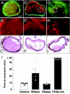
Fig. 1.
Fig. 1.
Association of extracellular RNA with thrombus formation and intervention with RNase in arterial thrombosis after vascular injury in vivo. Vascular injury of the mouse carotid artery was induced by local application of ferric chloride (A–I) in the absence (vehicle; A–C) or after pretreatment with RNase (D–F) or factor XIIa-inhibitor (G–I) as indicated (for details, see Materials and Methods). In all cases, before thrombus induction, an RNA-selective fluorescent green stain was administered (A, D, and G). Note the decoration of the thrombus by RNA in A and G, whereas pretreatment with RNase resulted in RNA hydrolysis and reduction of thrombus size (D) and pretreatment with FXIIa-inhibitor led to prevention of vessel occlusion without affecting RNA association with the thrombus (G). Postembedding staining against platelet glycoprotein Ib (red fluorescence in A, B, D, E, G, and H) indicates platelet-rich nature of thrombi. Counterstain for H&E is shown in C, F, and I for each group. (J) The time to thrombotic occlusion in the carotid artery downstream of the site of injury was assessed in vivo by video fluorescence microscopy in the absence (vehicle) or presence of RNase, DNase, or factor XIIa-inhibitor (FXIIa-Inh). Data represent mean ± SEM (n = 5–8 mice in each group). Circles represent results from individual experiments. ∗, P < 0.05 vs. vehicle and DNase (Mann–Whitney test). Representative images for each group are shown in SI Fig. 4.
Fig. 2.
Fig. 2.
Procoagulant activity of cell lysate and extracellular nucleic acids. (A) The procoagulant activity of particle free cell lysate prepared from cultured CHO cells was analyzed in a turbidity coagulation assay in the absence (blue diamonds) or presence of RNase (purple squares), DNase (yellow triangles), or antitissue factor (green crosses). Data represent mean ± SD, n = 3 of a representative experiment of three. (B) The procoagulant activity of cell lysate as well as of isolated RNA or DNA as indicated was tested in whole blood thrombelastography, whereby the time period (lag phase) until thrombus formation occurs and its extent is measured (amplitude) (Upper Left, Right, and Center). Parallel samples were pretreated with RNase A or DNase as indicated in each case, and representative experiments of six are shown. (C) Compared with kaolin (diamonds) different concentrations of RNA (squares) and DNA (triangles) were tested for procoagulant activity in pooled normal human plasma by using turbidity clot-lysis assay. Buffer control is indicated by the green circle, and nuclease-pretreated RNA and DNA are indicated by the red square and triangle, respectively. Data represent the mean ± SD (n = 3) of one representative experiment of three. (D) Anticoagulated normal human plasma was supplemented with 10 μg/ml each of RNA prepared from CHO cells, tRNA from yeast, total RNA from E. coli, single-stranded RNA from Qβ-phage, or buffer (control) as indicated, and coagulation was initiated by recalcification. Both untreated (gray) and RNase-pretreated (hatched) samples were compared. Data are converted from coagulation times into kaolin-equivalent units per ml, as explained in Materials and Methods and represent mean ± SD (n = 3) of three individual experiments. (E) Kaolin (10 ng/ml, open bar) and untreated and nuclease-pretreated RNA or DNA (10 μg/ml each) as indicated were incubated with citrated normal human plasma, and an increase in serine protease activity was followed by a chomogenic substrate (S-2288) assay. In comparison, recombinant tissue factor (TF) (0.8 μg/ml, hatched bar) was analyzed. Data represent mean ± SD (n = 3) of of one representative experiment of three. (F) Using the same experimental protocol, serine protease activity of RNA-supplemented citrated plasma was followed in the absence or presence of increasing doses of aprotinin (blue diamonds), corn trypsin inhibitor (green circles), factor XIa inhibitor J545 (purple squares), kallikrein inhibitor J626 (red circles), or hirudin (filled triangles). Data represent mean ± SD (n = 3) of one representative experiment of three.
Fig. 3.
Fig. 3.
Direct binding of nucleic acids to contact phase proteins and protease activation. (A) For detection of detergent-resistent RNA-protein complexes, radiolabeled HCV-RNA and blood coagulation factors as well as BSA were incubated and crosslinked by UV light, followed by RNase treatment, SDS/PAGE on 8% gels, and autoradiography (XII, factor XII; HK, kininogen; PK, prekallikrein; XI, factor XI; IX, factor IX; X, factor X; II, prothrombin; Fbg, fibrinogen). Molecular masses of 14C-labeled marker proteins are indicated along the left margin. (B) For electrophoretic mobility shift assays, radiolabeled HCV-RNA was incubated in the absence or presence of increasing doses (0.1, 0.3, 1, and 3 pmol) each of factor XI, factor X, or factor II, separated on non-denaturing 4% polyacrylamide gels and analyzed by autoradiography. The RNA-protein complexes and the main species of free RNA are indicated, and minor amounts of faster or slower migrating conformers of the unbound RNA are visible. (C) Factor XI alone (open bars), factor XII alone (hatched bars), or both (filled bars) were incubated in a zinc-containing buffer in the absence (−) or presence of RNA or DNA as indicated, and the extent of protease activation was followed by chromogenic substrate assay. (D) Similarly, factor XI alone (open bars), thrombin alone (hatched bars) or both together were analyzed for protease activation in the absence (−) or presence of RNA or DNA as indicated. (E) Shown are different reactions mixtures containing the following: 1, factor XII; 2, prekallikrein; 3, kininogen; 4, factor XII and prekallikrein; 5, factor XII, prekallikrein and kininogen were analyzed for protease activation in the absence (−) or the presence of RNA or DNA. All data represent mean ± SD (n = 3) of a representative experiment of three. (F) The activation of prekallikrein was followed in the presence of increasing doses of tRNA, and enzyme activity was registered by chromogenic substrate cleavage. Note the bell-shaped concentration curve, which is characteristic for a template-related mechanism of RNA as cofactor.

References

    1. Cines DB, Pollak ES, Buck CA, Loscalzo J, Zimmermann GA, McEver RP, Pober JS, Wicj TM, Konkle BA, Schwartz BS, et al. Blood. 1998;91:3527–3561. - PubMed
    1. Ruf W, Mueller BM. Thromb Haemost. 1999;82:175–182. - PubMed
    1. Mann KG, Butenas S, Brummel K. Arterioscler Thromb Vasc Biol. 2003;23:17–25. - PubMed
    1. Müller I, Klocke A, Alex M, Kotzsch M, Luther T, Morgenstern E, Zieseniss S, Zahler S, Preissner KT, Engelmann B. FASEB J. 2003;17:476–478. - PubMed
    1. Davie EW, Ratnoff OD. Science. 1964;145:1310–1312. - PubMed

Publication types

LinkOut - more resources