. 2007 Apr 23;96(8):1246-52.
doi: 10.1038/sj.bjc.6603700. Epub 2007 Apr 3.

Co-culturing human prostate carcinoma cells with hepatocytes leads to increased expression of E-cadherin

Affiliations

Co-culturing human prostate carcinoma cells with hepatocytes leads to increased expression of E-cadherin

C C Yates et al. Br J Cancer. .

Abstract

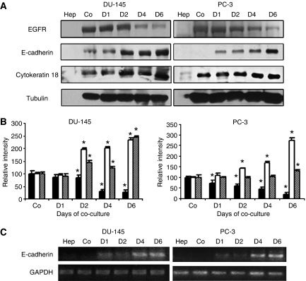
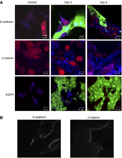
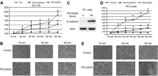
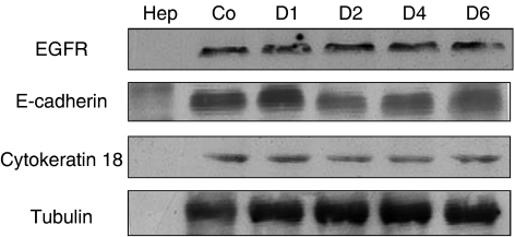
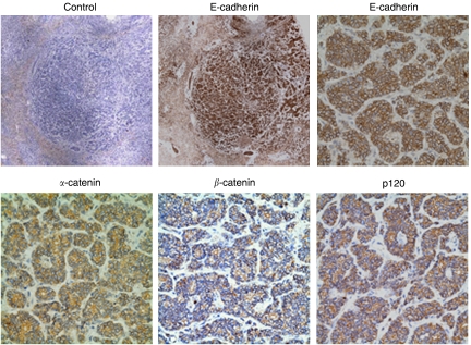
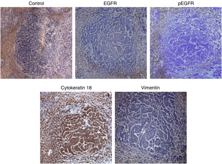
Metastasis is a multi-step process wherein tumour cells detach from the primary mass, migrate through barrier matrices, gain access to conduits to disseminate, and subsequently survive and proliferate in an ectopic site. During the initial invasion stage, prostate carcinoma cells undergo epithelial-mesenchymal-like transition with gain of autocrine signalling and loss of E-cadherin, hallmarks that appear to enable invasion and dissemination. However, some metastases express E-cadherin, and we found close connections between prostate carcinoma cells and hepatocytes in a liver microtissue bioreactor. We hypothesise that phenotypic plasticity occurs late in prostate cancer progression at the site of ectopic seeding. Immunofluorescence staining for E-cadherin in co-cultures of hepatocytes and DU-145 prostate cancer cells revealed E-cadherin upregulation at peripheral sites of contact by day 2 of co-culture; E-cadherin expression also increased in PC-3 cells in co-culture. These carcinoma cells bound to hepatocytes in an E-cadherin-dependent manner. Although the signals by which the hepatocytes elicited E-cadherin expression remain undetermined, it appeared related to downregulation of epidermal growth factor receptor (EGFR) signalling. Inhibition of autocrine EGFR signalling increased E-cadherin expression and cell-cell heterotypic adhesion; further, expression of a downregulation-resistant EGFR variant prevented E-cadherin upregulation. These findings were supported by finding E-cadherin and catenins but not activated EGFR in human prostate metastases to the liver. We conclude that the term epithelial-mesenchymal transition only summarises the transient downregulation of E-cadherin for invasion with re-expression of E-cadherin being a physiological consequence of metastatic seeding.


Figures

Figure 1
Figure 1
Co-culture of human prostate cancer cell with rat hepatocytes reversed E-cadherin expression. DU-145 or PC-3 (A) cells were co-cultured in the presence of primary rat hepatocytes over a 6-day period. Hepatocytes and single cultures were lysed before co-cultures. On days 1, 2, 4 and 6, co-culture lysates were immunoblotted with indicated human selective antibodies: anti-E-cadherin, anti-EGFR antibody, anti-cytokeratin 18 and anti-tubulin (as the loading control). (B) Densitometry of immunoblots in DU-145 and PC-3 cells co-cultures (▪) EGFR, (□) E-cadherin () and Cytokeratin 18; shown are the mean±s.d. of three blots with day 0 being at 100 (*P<0.05 from C0). Cellular levels of E-cadherin mRNA in DU-145 or PC-3 (C) cells were analysed by RT-PCR using GAPDH as a loading control. In A and B, Co (for control) are an equal number of prostate carcinoma cells incubated for 1 day in the absence of hepatocytes. The Hep (hepatocytes) were an equal number of hepatocytes as for the co-cultures. The first three lanes were all lysed at the same time. Shown are representative of at least three experiments.
Figure 2
Figure 2
Immunofluorescence of co-cultures show subcellular location of E-cadherin re-expression. (A) Immunofluorescence of DU-145 RFP (red) and GFP (green) primary rat hepatocytes were stained with human-specific anti-E-cadherin, pan-species anti-β-catenin or human-specific anti-EGFR antibody. Cy5 secondary antibody (blue) was used for respective primary antibodies. Note the gain of blue (E-cadherin) in the RFP/red prostate cells in the top and middle rows, and the loss of blue (EGFR) in the bottom row. (B) The blue channel only of the lower left inset in the E-cadherin and β-catenin staining on day 2 is shown in black and white to demonstrate the localisation of the human E-cadherin in the prostate carcinoma cells to the interface with the hepatocytes. In the middle row, the hepatocytes were from WT and not GFP rats, so as not to interfere with the antibody staining as the anti-β-catenin detected both human and rat. However, the presence of β-catenin upregulation in the DU-145 cells is noted by a violet color and the membranous pattern at the hepatocyte–prostate carcinoma cell interface. Shown are representative of at least three experiments.
Figure 3
Figure 3
Human prostate cancer cells form E-cadherin-mediated heterotypic interactions with hepatocytes. DU-145 (A and B) cells or PC-3 (CE) cells fluorescently labelled with Calcein A were incubated for 48 h with PD153035 or E-cadherin blocking antibody and seeded onto a monolayer of hepatocytes from non-GFP-expressing rats to analyse their ability to adhere. Cell binding was assessed by fluorescent intensity using a plate fluorometer and visually verified under a fluorescent light microscope. Y-axis is arbitrary fluorescent units. Data represent mean of three experiments performed in triplicate; s.e. *P<0.05. Shown are sample representative fields to show the bound tumour cells (converted to white dots) overlying the hepatocytes in B and E. (C) PC-3 cells were exposed to PD153035 for 48 h with a resultant upregulation of E-cadherin as shown by immunoblotting. This is similar to our previously published finding with DU145 cells (Yates et al, 2005).
Figure 4
Figure 4
DU-145 cells expressing a PKC transattenuation-resistant EGFR (A654) are resistant to hepatocyte-induced E-cadherin re-expression. DU-145 A654 cells co-culture lysates were immunoblotted with an antibody selective for human E-cadherin, EGFR, cytokeratin 18 or tubulin. The legend is as with Figure 1, Co (control) DU145 A654 cells and Hep (hepatocytes) only. Shown is one of two representative blot series.
Figure 5
Figure 5
Human prostate cancer metastases to liver show expression of cell–cell adhesion molecules. Formalin-fixed, paraffin-embedded tissues were obtained from two well-defined prostate adenocarcinomas with liver metastasis. Tissues were stained with indicated antibodies, Secondary antibody, anti-mouse only as the staining control (top left; 3700 μm2), anti-E-cadherin (top centre; 3700 μm2 and top right; 300 μm2), anti-α-catenin (bottom right; 300 μm2), anti-β-catenin (bottom centre; 300 μm2) and anti-p120 (bottom right; 300 μm2). Shown are representative of repeated stainings; the other metastasis presented similar findings.
Figure 6
Figure 6
Human prostate cancer metastases show reversion of metastatic markers. Tissues were stained with anti-rabbit (top left; 1400 μm2) anti-EGFR (top centre; 1400 μm2), anti-phosphotyrosyl-EGFR (activated EGFR) (top right; 1400 μm2), anti-vimentin (bottom left; 1400 μm2) and anti-cytokeratin 18 (bottom left; 1400 μm2). Shown are representative of repeated stainings; the other metastasis presented similar findings.

References

    1. Ackland ML, Newgreen DF, Fridman M, Waltham MC, Arvanitis A, Minichiello J, Price JT, Thompson EW (2003) Epidermal growth factor-induced epithelio–mesenchymal transition in human breast carcinoma cells. Lab Invest 83: 435–448 - PubMed
    1. Angelucci A, Gravina GL, Rucci N, Millimaggi D, Festuccia C, Muzi P, Teti A, Vicentini C, Bologna M (2006) Suppression of EGF-R signaling reduces the incidence of prostate cancer metastasis in nude mice. Endocr Relat Cancer 13: 197–210 - PubMed
    1. Bates RC, Mercurio AM (2005) The epithelial–mesenchymal transition (EMT) and colorectal cancer progression. Cancer Biol Ther 4: 365–370 - PubMed
    1. Brabletz T, Jung A, Reu S, Porzner M, Hlubek F, Kunz-Schughart LA, Knuechel R, Kirchner T (2001) Variable beta-catenin expression in colorectal cancers indicates tumor progression driven by the tumor environment. Proc Natl Acad Sci USA 98: 10356–10361 - PMC - PubMed
    1. Bryden AA, Hoyland JA, Freemont AJ, Clarke NW, Schembri-Wismayer D, George NJ (2002) E-cadherin and beta catenin are down-regulated in prostatic bone mestatases. BJU Intl 89: 400–403 - PubMed

Publication types