. 2006;1(3):1145-53.
doi: 10.1038/nprot.2006.173.

Production of chimeras by aggregation of embryonic stem cells with diploid or tetraploid mouse embryos

Affiliations

Production of chimeras by aggregation of embryonic stem cells with diploid or tetraploid mouse embryos

Guy S Eakin et al. Nat Protoc. 2006.

Abstract

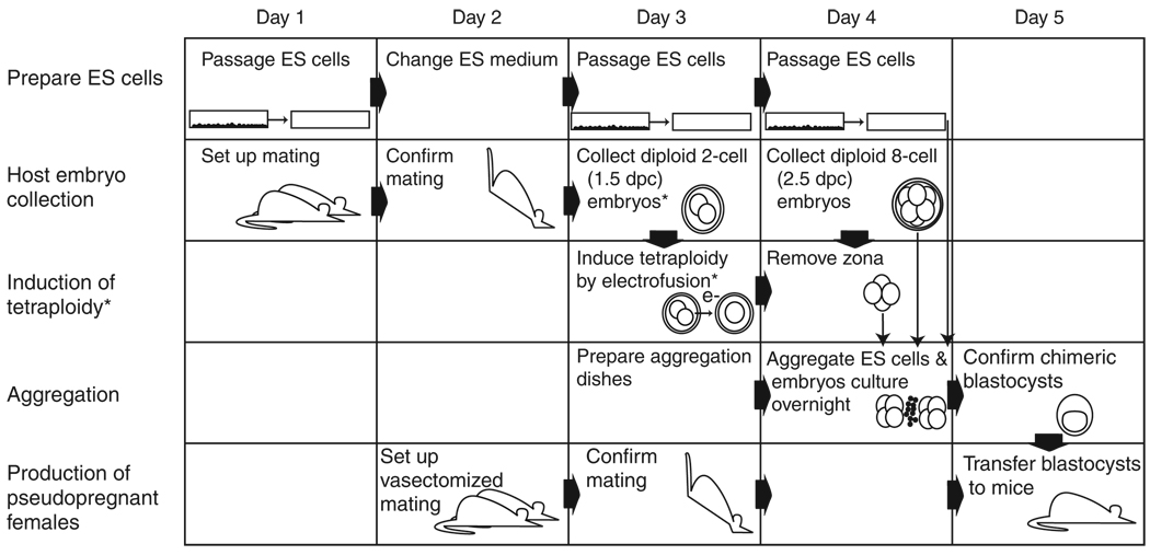
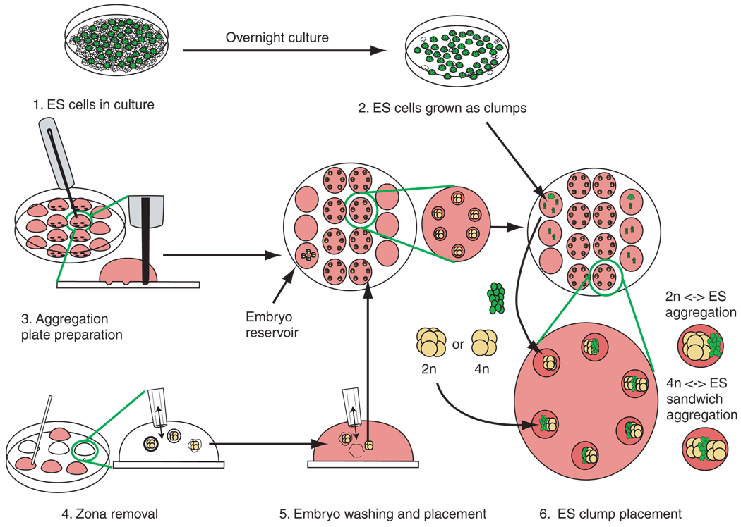
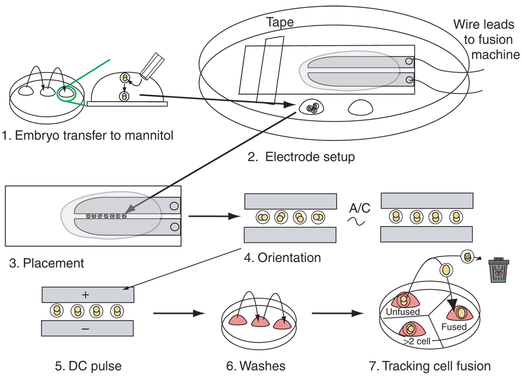
The production of mouse chimeras is a common step in the establishment of genetically modified animal strains. Chimeras also provide a powerful experimental tool for following cell behavior during both prenatal and postnatal development. This protocol outlines a simple and economical technique for the production of large numbers of mouse chimeras using traditional diploid morula<-->diploid embryonic stem (ES) cell aggregations. Additional steps are included to describe the procedures necessary to produce specialized tetraploid chimeras using tetraploid morula<-->diploid ES cell aggregations. This increasingly popular form of chimera produces embryos of nearly complete ES cell derivation that can be used to speed transgenic production or ask developmental questions. Using this protocol, mouse chimeras can be generated and transferred to pseudopregnant surrogate mothers in a 5-d period.


Figures

Figure 1
Figure 1
Timeline and integration of the protocols. The exact regimen for the preparation of ES cells varies with different cell lines. Usually, ES cell medium is changed daily, and cells are passaged on the second or third day once they reach 70% confluency. For the generation of aggregation chimeras, it is imperative that cells are passaged in such a way that they form clumps of cells that can be recovered from the dish after brief treatment with trypsin. Experiments with mice need to be carefully planned; matings to produce the embryos required for the aggregations and pseudopregnant females for the transfer of chimeric blastocysts need to be synchronized. For the routine production of diploid embryo ↔ diploid ES cell chimeras, embryos are recovered at the eight-cell stage at 2.5 dpc when they appear as uncompacted morulae. The zona is immediately removed, and the embryos are then used to set up the aggregation. Embryos can, however, also be recovered 1 or 2 d earlier at 1.5 or 0.5 dpc and cultured until they form morulae, at which time the zona is removed before aggregation setup. The same applies for embryos that are to be rendered tetraploid, which can be recovered at 0.5 dpc and cultured overnight until they reach the two-cell stage. Asterisks indicate the steps that are unique to the production of tetraploid (4n) embryo ↔ diploid ES cell chimeras.
Figure 2
Figure 2
Generalized scheme for the production of aggregation chimeras. The procedure can be divided into six parts. 1. ES cells are passaged to deplete feeders. 2. ES cells are lightly trypsinized to release small clumps. ES cell clumps are transferred to drops serving as ES reservoirs on the outer rows of the aggregation plate. 3. The aggregation plate routinely contains four rows of KSOM drops: two outer rows of three drops that serve as ES cell and embryo reservoirs and two inner rows of four drops that contain depressions required for the aggregation of embryos with ES cells. Once made, the drops are overlayed with mineral oil. Depression wells are then made in the central two rows of wells of the aggregation plate. We routinely make six depressions per drop. It is advisable that the depressions are made and the plate be equilibrated in an incubator before the addition of cells or embryos. 4. The zona pellucida of the embryos is dissolved in Acid Tyrode’s solution. It is important to keep pipetting the embryos, as they tend to become sticky and should not make prolonged contact with the plastic surface of the dish, otherwise they can be difficult to dislodge. 5. The embryos are then washed through M2 medium. Zona-free embryos are transferred to a reservoir drop on the aggregation plate. Single embryos are immediately transferred into each depression. This must be done quickly, as zona-free embryos will aggregate with each other if left in contact. If tetraploid chimeras are being set up, then half of the embryos should be left in a reservoir until the ES cell clumps have been added to the embryos in the depressions. 6. ES cell clumps are transferred from the reservoirs to the depression wells. Care must be taken that the ES cells are in physical contact with the embryos, otherwise the embryos may form blastocysts that will not have incorporated the ES cells. For tetraploid aggregations, a second embryo is then placed in the depression so that the ES cells are sandwiched between two embryos.
Figure 3
Figure 3
Protocol for generating tetraploid embryos by electrofusion. The procedures can be divided into seven parts. 1. Embryos are washed through mannitol solution, allowing them to sink to the bottom of the drop. 2. The electrode slide is placed (taped or clipped) to the lid of a 10-cm tissue culture dish. The embryos are transferred to a reservoir drop of mannitol placed on the plastic dish to the side of the electrode slide. 3. The embryos (generally 15–20 at a time) are lined up in single file between the electrodes. 4. Embryos are oriented with the interface between the two blastomeres perpendicular to the electrodes using an A/C orientation field. 5. The aligned embryos are pulsed with DC current to initiate blastomere fusion. 6. Immediately after pulsing, embryos are washed through sequential drops of M2 medium to remove any remaining mannitol. Embryos are then sequentially washed into KSOM medium. 7. They are then placed in a drop of KSOM in a dish containing three drops of KSOM. The drops are labeled ‘unfused’, ‘fused’ and ‘cleaved’ or ‘>2 cell’. The KSOM drops are covered in mineral oil, and the plate is placed in an incubator. The plate is removed from the incubator at 10- to 15-min intervals to monitor the process of fusion. As blastomeres fuse, individual embryos are immediately transferred from the unfused drop to the fused drop. Unhealthy or lysing embryos are discarded. The drop labeled ‘>2 cell’ can be used for placing older, unfused (diploid) embryos. These embryos will begin to cleave soon after pulsing and should immediately be removed from the pool of fusing embryos so as not to be confused with embryos that may have fused and then cleaved.
Figure 4
Figure 4
Morphology of embryos throughout the procedure. (a,b) Development of zona-free embryos in depression wells: zona-free morula (a) and zona-free blastocyst after 24 h in culture (b). White arrow, depression well; black arrow, inner cell mass of blastocyst. (ce) Induction of tetraploidy by electrofusion. Also see Supplementary Video 1. Two-cell diploid embryo before fusion (c). Initial stages of fusion as plasma membrane breaks down (d). Fusion is completed, which results in a one-cell tetraploid embryo (e). (fh) In vitro development of tetraploid embryos. Two-cell tetraploid embryo 12 h after fusion (f). Four-cell tetraploid embryo 24 h after fusion (g). Tetraploid blastocyst 48 h after fusion (h). A series of images that shows the progression of both a diploid embryo ↔ diploid ES cell aggregation and a tetraploid embryo ↔ diploid ES cell aggregation, where the ES cells are expressing a GFP transgene, has been reported.

References

    1. Bradley A, Evans M, Kaufman MH, Robertson E. Formation of germ-line chimeras from embryo-derived teratocarcinoma cell lines. Nature. 1984;309:255–256. - PubMed
    1. Thompson S, Clarke AR, Pow AM, Hooper ML, Melton DW. Germ line transmission and expression of a corrected HPRT gene produced by gene targeting in embryonic stem cells. Cell. 1989;56:313–321. - PubMed
    1. Tam PP, Rossant J. Mouse embryonic chimeras: tools for studying mammalian development. Development. 2003;130:6155–6163. - PubMed
    1. Kunath T, et al. Imprinted X-inactivation in extra-embryonic endoderm cell lines from mouse blastocysts. Development. 2005;132:1649–1661. - PubMed
    1. Tanaka S, Kunath T, Hadjantonakis AK, Nagy A, Rossant J. Promotion of trophoblast stem cell proliferation by FGF4. Science. 1998;282:2072–2075. - PubMed

LinkOut - more resources